Rebamipide ameliorates atherosclerosis by controlling lipid metabolism and inflammation
- PMID: 28241014
- PMCID: PMC5328247
- DOI: 10.1371/journal.pone.0171674
Rebamipide ameliorates atherosclerosis by controlling lipid metabolism and inflammation
Abstract
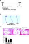
The oral administration of rebamipide decreased plaque formation in atherosclerotic lesions as well as the markers of metabolic disorder in ApoE-deficient mice with atherosclerosis. Pro-inflammatory cytokines were also suppressed by rebamapide. In addition, the population of Th17 was decreased, whereas Treg was increased in the spleen of rebamipide-treated ApoE deficient mice. Rebamipide also ameliorated the severity of obese arthritis and has the capability to reduce the development of atherosclerosis by controlling the balance between Th17 and Treg cells. Thus, rebamipide could be a therapeutic agent to improve the progression of inflammation in metabolic diseases.
Conflict of interest statement
Figures


Similar articles
-
Rebamipide suppresses collagen-induced arthritis through reciprocal regulation of th17/treg cell differentiation and heme oxygenase 1 induction.Arthritis Rheumatol. 2014 Apr;66(4):874-85. doi: 10.1002/art.38310. Arthritis Rheumatol. 2014. PMID: 24757140
-
Rebamipide prevents peripheral arthritis and intestinal inflammation by reciprocally regulating Th17/Treg cell imbalance in mice with curdlan-induced spondyloarthritis.J Transl Med. 2016 Jun 27;14(1):190. doi: 10.1186/s12967-016-0942-5. J Transl Med. 2016. PMID: 27350608 Free PMC article.
-
TSLPR deficiency attenuates atherosclerotic lesion development associated with the inhibition of TH17 cells and the promotion of regulator T cells in ApoE-deficient mice.J Mol Cell Cardiol. 2014 Nov;76:33-45. doi: 10.1016/j.yjmcc.2014.07.003. Epub 2014 Aug 10. J Mol Cell Cardiol. 2014. PMID: 25117469
-
Diet and atherosclerosis in apolipoprotein E-deficient mice.Biosci Biotechnol Biochem. 2011;75(6):1023-35. doi: 10.1271/bbb.110059. Epub 2011 Jun 13. Biosci Biotechnol Biochem. 2011. PMID: 21670535 Review.
-
Interleukin-17 as a factor linking the pathogenesis of psoriasis with metabolic disorders.Int J Dermatol. 2017 Mar;56(3):260-268. doi: 10.1111/ijd.13420. Epub 2016 Oct 1. Int J Dermatol. 2017. PMID: 27696392 Review.
Cited by
-
Rebamipide treatment ameliorates obesity phenotype by regulation of immune cells and adipocytes.PLoS One. 2022 Dec 27;17(12):e0277692. doi: 10.1371/journal.pone.0277692. eCollection 2022. PLoS One. 2022. PMID: 36574392 Free PMC article.
References
-
- Zhou X. CD4+ T cells in atherosclerosis. Biomed Pharmacother. 2003;57(7):287–91. - PubMed
-
- Jonasson L, Holm J, Skalli O, Bondjers G, Hansson GK. Regional accumulations of T cells, macrophages, and smooth muscle cells in the human atherosclerotic plaque. Arteriosclerosis. 1986;6(2):131–8. - PubMed
-
- Zhou X, Nicoletti A, Elhage R, Hansson GK. Transfer of CD4(+) T cells aggravates atherosclerosis in immunodeficient apolipoprotein E knockout mice. Circulation. 2000;102(24):2919–22. - PubMed
MeSH terms
Substances
LinkOut - more resources
Full Text Sources
Other Literature Sources
Medical
Molecular Biology Databases
Miscellaneous

