

Restored vision in a young dog following corticosteroid treatment of presumptive hypophysitis
- PMID: 28241874
- PMCID: PMC5330113
- DOI: 10.1186/s12917-017-0983-x
Restored vision in a young dog following corticosteroid treatment of presumptive hypophysitis
Abstract
Background: Hypophysitis is an umbrella term for a group of disorders involving inflammation of the pituitary gland. A rare occurrence in humans, hypophysitis can produce a range of clinical signs including (but not limited to) visual deficits and diabetes insipidus. Only five cases of canine hypophysitis exist in the literature, all presenting in mature dogs with no visual deficits and a grave outcome. This case report describes the clinical and advanced imaging features of blindness-inducing presumptive hypophysitis in a dog, which rapidly resolved with medical management.
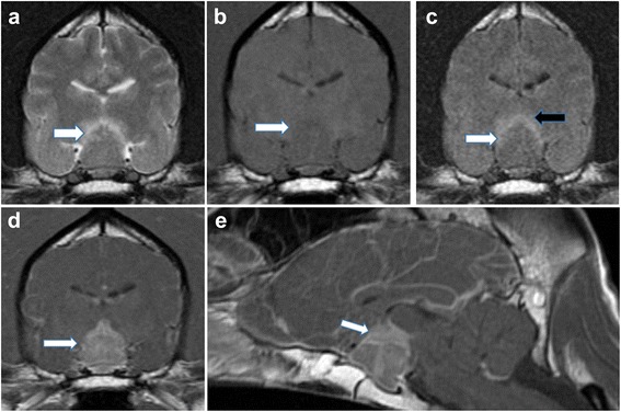
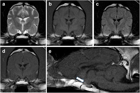
Case presentation: A 1-year-and-seven-month-old neutered male Standard Poodle presented with subacute blindness, ataxia, and polyuria/polydipsia (PUPD). Magnetic resonance imaging (MRI) detected a contrast-enhancing pituitary mass with perilesional oedema compromising the optic chiasm. Suspecting neoplasia, anti-inflammatory corticosteroid was commenced prior to radiation therapy planning. Complete resolution of neurological and visual deficits occurred within 12 days of starting steroid treatment. Repeated advanced imaging indicated macroscopic resolution of the lesion. An extended thyroid panel with insulin-like growth factor-1 analysis supported a diagnosis of hypophysitis. Resolution of PUPD was achieved with tapering courses of prednisolone and desmopressin; the dog has since been clinically normal for 14 months and treatment-free for 11 months.
Conclusions: To the authors' knowledge, this is the first instance in which a canine pituitary mass has demonstrated long-term resolution with palliative medical treatment alone, alongside reversal of associated blindness and presumptive diabetes insipidus. We suspect this lesion to be a form of hypophysitis, which should be included among differential diagnoses for pituitary masses, and for subacute blindness in dogs. Where possible, we advocate biopsy-confirmation of hypophysitis prior to timely intervention with anti-inflammatory treatment.
Keywords: Central blindness; Hypophysitis; Insulin-like growth factor-1; Magnetic resonance imaging; Pituitary tumour; Standard Poodle.
Figures
Similar articles
-
Hypophysitis, Panhypopituitarism, and Hypothalamitis in a Scottish Terrier Dog.J Vet Intern Med. 2017 Sep;31(5):1527-1532. doi: 10.1111/jvim.14790. Epub 2017 Jul 26. J Vet Intern Med. 2017. PMID: 28745808 Free PMC article.
-
IgG4-related hypophysitis diagnosed by retroperitoneal mass biopsy in a patient presenting with abducens nerve palsy: A case report (CARE-compliant article).Medicine (Baltimore). 2020 Oct 2;99(40):e22484. doi: 10.1097/MD.0000000000022484. Medicine (Baltimore). 2020. PMID: 33019443 Free PMC article.
-
AVP deficiency (central diabetes insipidus) following immunization with anti-COVID-19 BNT162b2 Comirnaty vaccine in adolescents: A case report.Front Endocrinol (Lausanne). 2023 Apr 18;14:1166953. doi: 10.3389/fendo.2023.1166953. eCollection 2023. Front Endocrinol (Lausanne). 2023. PMID: 37143723 Free PMC article.
-
Bilateral Optic Neuritis and Hypophysitis With Diabetes Insipidus 1 Month After COVID-19 mRNA Vaccine: Case Report and Literature Review.J Investig Med High Impact Case Rep. 2023 Jan-Dec;11:23247096231186046. doi: 10.1177/23247096231186046. J Investig Med High Impact Case Rep. 2023. PMID: 37431875 Free PMC article. Review.
-
[Lymphocytic infundibulo-hypophysitis with diabetes insipidus as a new clinical entity: a case report and review of the literature].No Shinkei Geka. 1997 Feb;25(2):169-75. No Shinkei Geka. 1997. PMID: 9027895 Review. Japanese.
Cited by
-
Hypophysitis, Panhypopituitarism, and Hypothalamitis in a Scottish Terrier Dog.J Vet Intern Med. 2017 Sep;31(5):1527-1532. doi: 10.1111/jvim.14790. Epub 2017 Jul 26. J Vet Intern Med. 2017. PMID: 28745808 Free PMC article.
References
-
- Wisner ER, Zwingenberger AL:. Section 2.2.9 Sellar and Parasellar Region. In: Wisner ER, Zwingenberger AL. Atlas of Small Animal CT and MRI. Hoboken: John Wiley & Sons; 2015. p. 244–263.
-
- Withrow SJ, Vail DM, Page RL. Withrow & MacEwen’s Small Animal Clinical Oncology. Fifth Edition. Philadelphia: Saunders, Elsevier Inc; 2013. pp. 189–190 and 504–505.
-
- Oliveira M, Polledo L, Adamany J, Wessmann A, Graham P, Dhumeaux M, et al. Hypophysitis in a Scottish Terrier with associated panhypopituitarism and hypothalamitis mimicking a pituitary gland neoplasia. Proceedings of the 29th Annual Symposium of the European Society of Veterinary Neurology and European College of Veterinary Neurology. 2016 September 15 to 17, Edinburgh, UK, p. 41–42.
Publication types
MeSH terms
Substances
Grants and funding
LinkOut - more resources
Full Text Sources
Other Literature Sources
Medical
