

Targeting the programmed death-1 pathway in lymphoid neoplasms
- PMID: 28242522
- PMCID: PMC5815314
- DOI: 10.1016/j.ctrv.2017.01.009
Targeting the programmed death-1 pathway in lymphoid neoplasms
Abstract
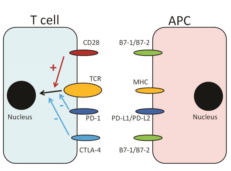
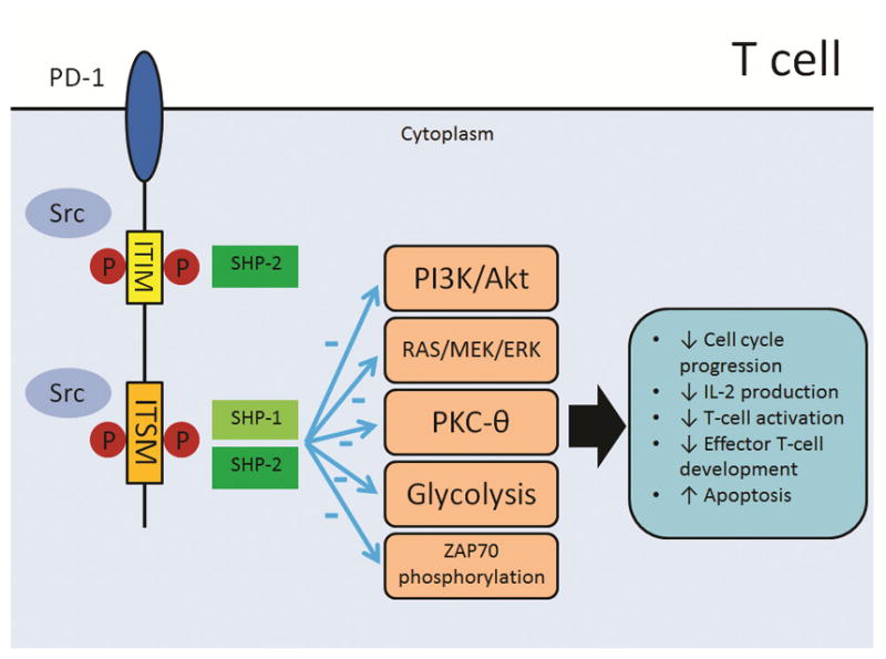
Programmed death-1 (PD-1) is a co-inhibitory molecule and is seen in CD4+ and CD8+ T cells. Upon binding to its ligands, programmed death ligand-1 (PD-L1) and -2 (PD-L2), PD-1 negatively regulates interleukin 2 (IL-2) production and T cell proliferation. Activated effector T-cells, which kill cancer cells, can be affected by PD-1 signaling in some lymphoid neoplasm that express PD-L1 or PD-L2. PD-L1 expression in tumor cells can be induced by extrinsic signal (i.e. interferon gamma) or intrinsic signals, such as genetic aberrations involving 9p24.1, latent Epstein-Barr virus infection, PD-L1 3'- untranslated region disruptions, and activated Janus kinase/signal transducer and activator of transcription (JAK/STAT) pathway. Anti-PD-1 therapy improves the overall response rate to treatment in patients with lymphoid neoplasms, particularly relapsed/refractory classical Hodgkin lymphoma. Inspired by their success in treating patients with classical Hodgkin lymphoma, medical practitioners have expanded PD-1 therapy, given as a single therapy or in combination with other drugs, to patients with other types of lymphoma. In this review, current clinical trials with anti-PD-1 or anti-PD-L1 drugs are summarized. The results of numerous clinical trials will broaden our understanding of PD-1 pathway and shall expand the list of patients who will get benefit from these agents including those who suffer from lymphoid neoplasms.
Keywords: Immune checkpoint; Lymphoid neoplasms; PD-1; PD-L1; PD-L2.
Copyright © 2017 Elsevier Ltd. All rights reserved.
Figures
Similar articles
-
Soluble programmed cell death protein 1 (sPD-1) and the soluble programmed cell death ligands 1 and 2 (sPD-L1 and sPD-L2) in lymphoid malignancies.Eur J Haematol. 2021 Jul;107(1):81-91. doi: 10.1111/ejh.13621. Epub 2021 Apr 12. Eur J Haematol. 2021. PMID: 33721375
-
PD-1/PD-L1 Pathway and Its Blockade in Patients with Classic Hodgkin Lymphoma and Non-Hodgkin Large-Cell Lymphomas.Curr Hematol Malig Rep. 2020 Aug;15(4):372-381. doi: 10.1007/s11899-020-00589-y. Curr Hematol Malig Rep. 2020. PMID: 32394185 Review.
-
Nivolumab in the Treatment of Hodgkin Lymphoma.Clin Cancer Res. 2017 Apr 1;23(7):1623-1626. doi: 10.1158/1078-0432.CCR-16-1387. Epub 2016 Nov 23. Clin Cancer Res. 2017. PMID: 27881581 Review.
-
Structural Biology of the Immune Checkpoint Receptor PD-1 and Its Ligands PD-L1/PD-L2.Structure. 2017 Aug 1;25(8):1163-1174. doi: 10.1016/j.str.2017.06.011. Structure. 2017. PMID: 28768162 Review.
-
Structure and interactions of the human programmed cell death 1 receptor.J Biol Chem. 2013 Apr 26;288(17):11771-85. doi: 10.1074/jbc.M112.448126. Epub 2013 Feb 15. J Biol Chem. 2013. PMID: 23417675 Free PMC article.
Cited by
-
A proportion of CD4+ T cells from patients with chronic Chagas disease undergo a dysfunctional process, which is partially reversed by benznidazole treatment.PLoS Negl Trop Dis. 2021 Feb 4;15(2):e0009059. doi: 10.1371/journal.pntd.0009059. eCollection 2021 Feb. PLoS Negl Trop Dis. 2021. PMID: 33539379 Free PMC article.
-
New insights into the important roles of tumor cell-intrinsic PD-1.Int J Biol Sci. 2021 Jun 16;17(10):2537-2547. doi: 10.7150/ijbs.60114. eCollection 2021. Int J Biol Sci. 2021. PMID: 34326692 Free PMC article. Review.
-
Immune checkpoint inhibitors and cellular treatment for lymphoma immunotherapy.Clin Exp Immunol. 2021 Jul;205(1):1-11. doi: 10.1111/cei.13592. Epub 2021 Mar 28. Clin Exp Immunol. 2021. PMID: 33675535 Free PMC article. Review.
-
Imaging diagnosis and efficacy monitoring by [89Zr]Zr-DFO-KN035 immunoPET in patients with PD-L1-positive solid malignancies.Theranostics. 2024 Jan 1;14(1):392-405. doi: 10.7150/thno.87243. eCollection 2024. Theranostics. 2024. PMID: 38164149 Free PMC article.
-
CAR-T cell combination therapies in hematologic malignancies.Exp Hematol Oncol. 2024 Jul 18;13(1):69. doi: 10.1186/s40164-024-00536-0. Exp Hematol Oncol. 2024. PMID: 39026380 Free PMC article. Review.
References
-
- Bretscher P, Cohn M. A theory of self-nonself discrimination. Science. 1970;169:1042–9. - PubMed
-
- Coyle AJ, Gutierrez-Ramos JC. The expanding B7 superfamily: increasing complexity in costimulatory signals regulating T cell function. Nat Immunol. 2001;2:203–9. - PubMed
-
- Brunet JF, Denizot F, Luciani MF, Roux-Dosseto M, Suzan M, Mattei MG, et al. A new member of the immunoglobulin superfamily--CTLA-4. Nature. 1987;328:267–70. - PubMed
-
- Waterhouse P, Penninger JM, Timms E, Wakeham A, Shahinian A, Lee KP, et al. Lymphoproliferative disorders with early lethality in mice deficient in Ctla-4. Science. 1995;270:985–8. - PubMed
Publication types
MeSH terms
Substances
Grants and funding
LinkOut - more resources
Full Text Sources
Other Literature Sources
Medical
Research Materials
