Review
. 2017 Aug:47:24-33.
doi: 10.1016/j.ceb.2017.02.005. Epub 2017 Feb 24.

Viral rewiring of cellular lipid metabolism to create membranous replication compartments

Affiliations
Review

Viral rewiring of cellular lipid metabolism to create membranous replication compartments

Jeroen Rpm Strating et al. Curr Opin Cell Biol. 2017 Aug.

Abstract

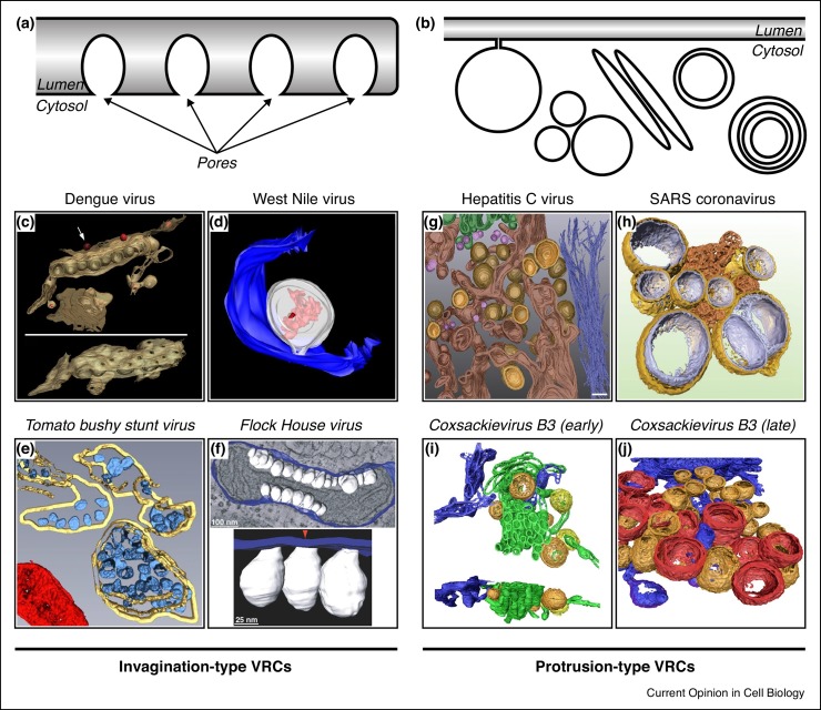
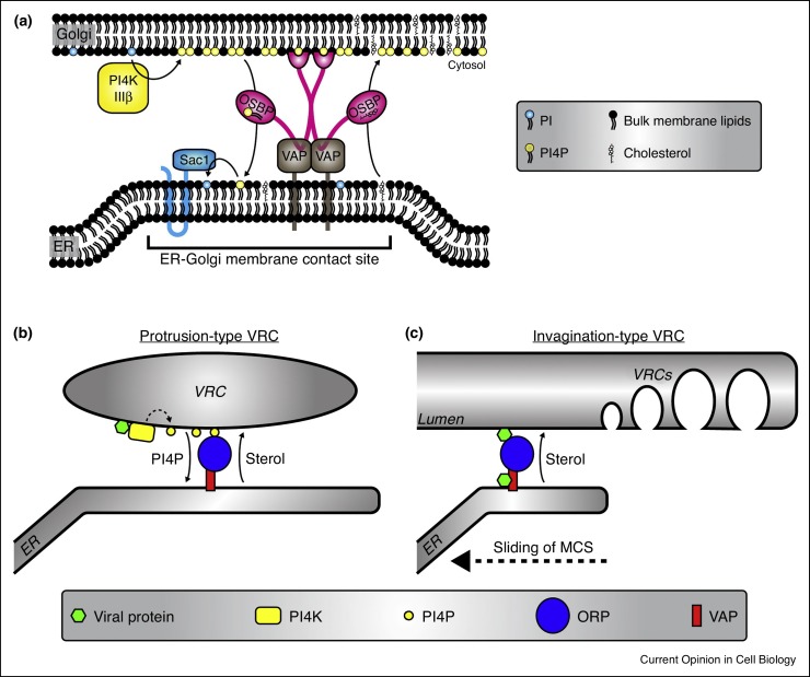
Positive-strand RNA (+RNA) viruses (e.g. poliovirus, hepatitis C virus, dengue virus, SARS-coronavirus) remodel cellular membranes to form so-called viral replication compartments (VRCs), which are the sites where viral RNA genome replication takes place. To induce VRC formation, these viruses extensively rewire lipid metabolism. Disparate viruses have many commonalities as well as disparities in their interactions with the host lipidome and accumulate specific sets of lipids (sterols, glycerophospholipids, sphingolipids) at their VRCs. Recent years have seen an upsurge in studies investigating the role of lipids in +RNA virus replication, in particular of sterols, and uncovered that membrane contact sites and lipid transfer proteins are hijacked by viruses and play pivotal roles in VRC formation.


Figures

Figure 1
Figure 1
Overview of different VRC morphologies. (a–b) Schematic overview of (a) invagination-type VRCs and (b) protrusion-type VRCs ([networks of] single- and double-membrane vesicles, multilamellar vesicles, tubules). (c–f) Three-dimensional reconstructions of invagination-type VRCs from various viruses. (c) VRCs of dengue virus in ER membranes. The tilted bottom panel is rotated by 90° to highlight the pores in the ER membrane that connect the VRC interior to the cytoplasm. (d) VRC (white) of West Nile virus in the lumen of the ER (blue). Viral RNA in the VRC lumen is displayed in red. (e) VRCs (blue) of tomato bushy stunt virus in the lumen of the peroxisome (yellow). A mitochondrion is shown in red. (f) VRCs (white) of Flock House virus in the intermembrane space of a mitochondrion connected to the outer mitochondrial membrane (blue). (g–j) Three-dimensional reconstructions of protrusion-type VRCs from various viruses. (g) Early stage of hepatitis C virus VRCs showing single-membrane (pink) and double-membrane (yellow inner membrane, light brown outer membrane) structures interspersed with ER membranes (dark brown). Golgi apparatus is shown in green, intermediate filaments are coloured dark blue. (h) Double-membrane vesicle VRCs of SARS-coronavirus (outer membrane in gold, inner membrane in silver) connected to so-called convoluted membranes (bronze). (i) Early stage tubular (green) and vesicular (orange, yellow) VRCs of the enterovirus coxsackievirus B3. ER is depicted in blue. (j) Late-stage VRCs of coxsackievirus B3 showing double-membrane vesicles (orange) and multilamellar vesicles (red). C has been reprinted from Ref. [79] with permission from Elsevier, D has been reproduced from Ref. [80] with permission from American Society for Microbiology, and E has been reproduced with permission from Journal of Cell Science from Ref. [81]. f–j are reproduced from open access publications [82] (f), [7] (g), [83] (h) and [5] (i, j).
Figure 2
Figure 2
Schematic depiction of cholesterol shuttling at MCSs. (a) Schematic depiction of OSBP-mediated cholesterol shuttling at ER-Golgi MCSs (based upon the model presented in [12••]). PI4KIIIβ produces PI4P lipids at the Golgi. PI4P serves as a docking site for OSBP dimers. VAP-A/B transmembrane proteins link OSBP to the ER. OSBP transports cholesterol against the concentration gradient from ER to Golgi. A counterflux of PI4P along the concentration gradient provides the driving force for cholesterol transport. In the ER, Sac1 hydrolyses PI4P into PI to keep the PI4P gradient intact. (b) Model of cholesterol shuttling at ER-VRC MCSs as proposed for enteroviruses, cardioviruses and HCV (recently reviewed in [84]). Viral proteins (i.e. enterovirus 3A, cardiovirus 3A or HCV NS5A) recruit a PI4K (i.e. PI4KIIIβ for enteroviruses, PI4KIIIα for HCV and cardioviruses) to enrich the VRC membranes in PI4P lipids (dotted arrow). Reminiscent of the physiological situation at the Golgi, the PI4P lipids anchor ORPs (in this case OSBP) to the VRCs and drive and OSBP-mediated cholesterol accumulation. (c) Model of cholesterol transport to invagination-type VRCs as proposed for TBSV [38••]. The viral protein p33 recruits ORPs (in this case Osh3, Osh5, Osh6 and Osh7) to the MCS between ER and the peroxisomes, while p33 also binds VAP at the ER. The ORPs mediate cholesterol accumulation at the peroxisome. Of note, Osh6 and Osh7 were shown to exchange PS instead of cholesterol for PI4P [85], suggesting that also PS may be shuttled to peroxisomes through the ER-peroxisome MCS. It has been hypothesised that the MCS slides along the surface of the peroxisome and that cholesterol accumulation primes the membrane for VRC formation in the wake of the sliding MCS [39].

Similar articles

Cited by

References

    1. Miller S., Krijnse-Locker J. Modification of intracellular membrane structures for virus replication. Nat Rev Microbiol. 2008;6:363–374. - PMC - PubMed
    1. Romero-Brey I., Bartenschlager R. Membranous replication factories induced by plus-strand RNA viruses. Viruses. 2014;6:2826–2857. - PMC - PubMed
    1. Bienz K., Egger D., Pfister T., Troxler M. Structural and functional characterization of the poliovirus replication complex. J Virol. 1992;66:2740–2747. - PMC - PubMed
    1. Egger D., Pasamontes L., Bolten R., Boyko V., Bienz K. Reversible dissociation of the poliovirus replication complex: functions and interactions of its components in viral RNA synthesis. J Virol. 1996;70:8675–8683. - PMC - PubMed
    1. Limpens R.W.A.L., van der Schaar H.M., Kumar D., Koster A.J., Snijder E.J., van Kuppeveld F.J.M., Bárcena M. The transformation of enterovirus replication structures: a three-dimensional study of single- and double-membrane compartments. mBio. 2011;2:e00111–e00166. - PMC - PubMed