. 2017 Apr 15;144(8):1477-1483.
doi: 10.1242/dev.143701. Epub 2017 Feb 27.

Stomach curvature is generated by left-right asymmetric gut morphogenesis

Affiliations

Stomach curvature is generated by left-right asymmetric gut morphogenesis

Adam Davis et al. Development. .

Abstract

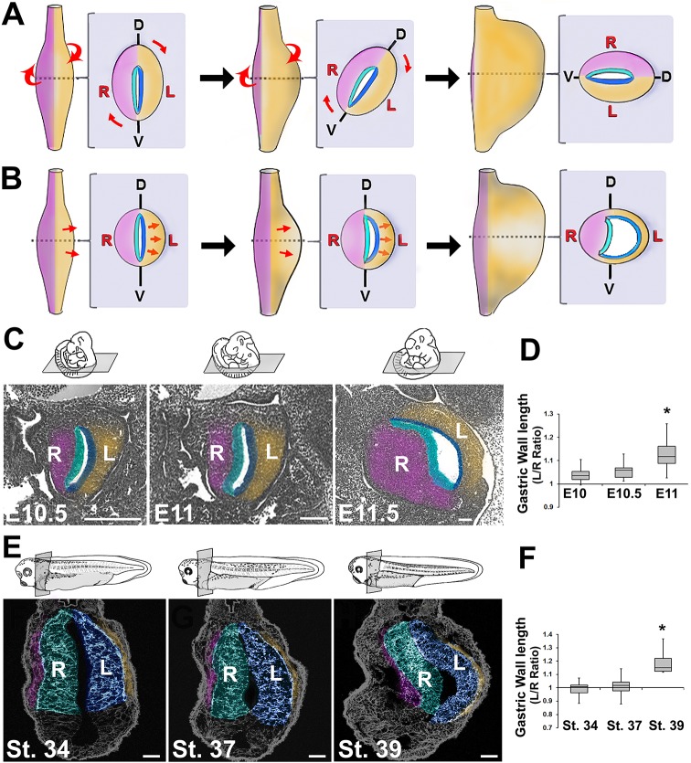
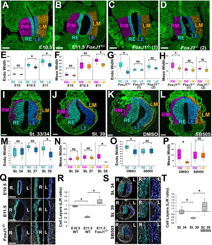
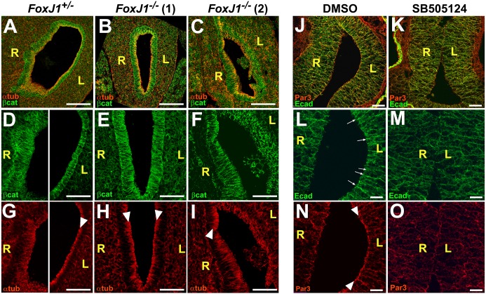
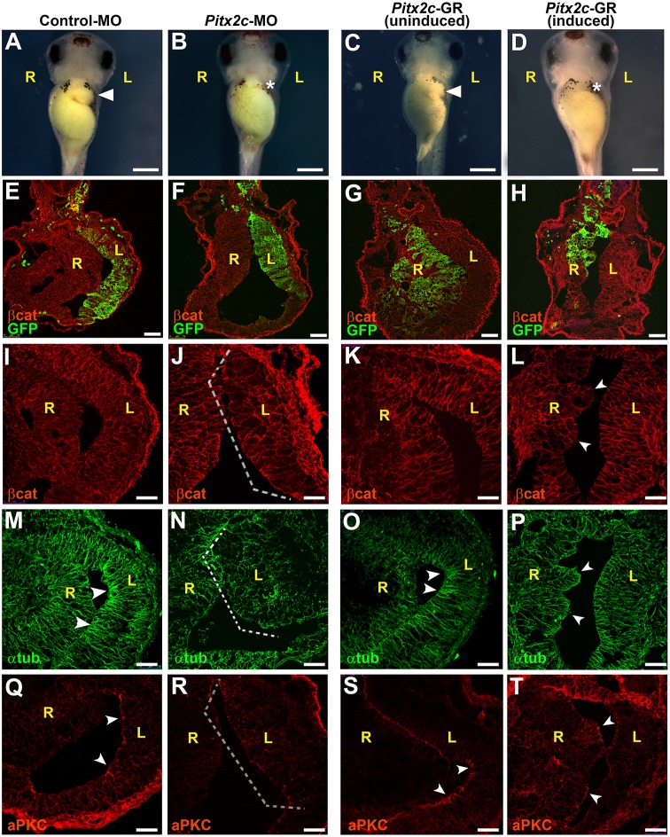
Left-right (LR) asymmetry is a fundamental feature of internal anatomy, yet the emergence of morphological asymmetry remains one of the least understood phases of organogenesis. Asymmetric rotation of the intestine is directed by forces outside the gut, but the morphogenetic events that generate anatomical asymmetry in other regions of the digestive tract remain unknown. Here, we show in mouse and Xenopus that the mechanisms that drive the curvature of the stomach are intrinsic to the gut tube itself. The left wall of the primitive stomach expands more than the right wall, as the left epithelium becomes more polarized and undergoes radial rearrangement. These asymmetries exist across several species, and are dependent on LR patterning genes, including Foxj1, Nodal and Pitx2 Our findings have implications for how LR patterning manifests distinct types of morphological asymmetries in different contexts.

Keywords: Asymmetry; Gut; Left-right; Morphogenesis; Mouse; Pitx2; Stomach; Xenopus.


Conflict of interest statement

Competing interests

The authors declare no competing or financial interests.

Figures

Fig. 1.
Fig. 1.
Early stomach undergoes leftward expansion. The rotation model (A) posits that the embryonic stomach (shown in ventral views and cross-sections at successive stages) rotates around its longitudinal axis, shifting its dorsal face leftward. An alternative model (B) theorizes that the left wall expands more than the right. Sections of E10.5, E11 and E11.5 mouse embryos (C) or stage 34, 37 and 39 frog embryos (E) reveal the leftward expansion of the early stomach. The left/right ratio of the lengths of the stomach walls becomes significantly greater than 1 in mouse by E11 (D) and in frog by stage 39 (F); *P<0.05. Sections in C and E are false-colored to match diagrams in A and B, highlighting layers of the stomach: right mesoderm, pink; right endoderm, teal; left endoderm, blue; left mesoderm, gold. In all sections, dorsal is upwards and the left side of animal is on right side of image. D, dorsal; V, ventral; L, left; R, right. Scale bars: 500 µm in C (E11.5, 150 µm); 75 µM in E.
Fig. 2.
Fig. 2.
Asymmetries in tissue architecture are regulated by left-right patterning. Sections through mouse (A-D) or frog (I-L) stomachs were stained for β-catenin (A-D) or integrin (I-L) (green) and false color-coded as in Fig. 1 (RE, right endoderm; LE, left endoderm; RM, right mesoderm; LM, left mesoderm). The widths of the endoderm (Endo) and mesoderm (Meso) are significantly different by E10.5-11 in mouse (E,F) and stage 39 in frog (M,N). In Foxj1+/− controls (E11.5), the lumen expands leftward and left-right differences in tissue width are evident (B); however, in Foxj1−/− mutants, the normal leftward expansion of the stomach is perturbed (C,D), and left-right differences in tissue width are eliminated (G,H). Likewise, frog embryos exposed to DMSO show normal leftward expansion of the stomach lumen (K); this is eliminated in embryos exposed to a Nodal inhibitor (SB505124; L), as are normal left-right differences in widths of endoderm and mesoderm (O,P). Nuclear staining reveals asymmetry in the number of endoderm cell layers in the right (teal) versus left (blue) stomach walls by E11.5 in mouse (Q,R) and stage 39 in frog (S,T); this asymmetry is perturbed in Foxj1−/− mutants (Q,R) and in frog embryos exposed to SB505124 (S,T). Scale bars: 100 µm in A-D,Q; 75 µm in I-L,S. *P<0.01; NS, not significant.
Fig. 3.
Fig. 3.
Asymmetries in stomach epithelial polarity are regulated by left-right signaling. Stomach sections immunostained for α-tubulin (αtub, red) and β-catenin (β-cat, green) in the E11.5 mouse (A-I), or Par3 (red) and E-cadherin (Ecad, green) in the stage 39 frog (J-O), reveal left-right differences in endoderm morphogenesis. In the posterior stomach of Foxj1+/− mice, right endoderm is irregular, whereas left endoderm is more organized (D) and highly polarized, as indicated by tight apical concentration of α-tubulin (αtub, red; arrowhead, G). Both walls of Foxj1−/− mutant embryo [Foxj1−/−(1), B,E,H], comprise polarized cells with tight apical concentration of αtub (arrowheads, H); both sides resemble control left epithelium. In another mutant [Foxj1−/−(2), C,F,I] both sides are irregular and resemble control right epithelium, although sub-regions exhibit greater organization and polarity (arrowhead, I). In control (DMSO) frog embryos (J,L,N), left endoderm is more polarized than right, as indicated by enrichment of E-cadherin (Ecad; green) at apical adherens junctions (arrows, L), and the concentration of the apical polarity marker Par3 at the lumenal surface (red; arrowheads, N). By contrast, SB505124 (K,M,O) results in right isomerism, with left endoderm resembling right endoderm, as indicated by absence of apical enrichment of E-cadherin (M) and Par3 (O). L, left; R, right. Scale bars: 100 µm in A-C; 50 µm in D-K; 25 µm in L-O. (See Fig. S4 for higher magnification images.)
Fig. 4.
Fig. 4.
Pitx2c controls epithelial morphogenesis in left stomach wall. Frog embryos were injected with control morpholino (control-MO; A,E,I,M,Q) or Pitx2c-MO (B,F,J,N,R) targeted to the left side of the stomach, or injected with Pitx2c-GR mRNA (C,G,K,O,S and D,H,L,P,T) targeted to the right side. (See Fig. S6A,B for morpholino validation.) In injected embryos (A-D), the greater curvature of the stomach at stage 42 is indicated by an arrowhead (A,C); absence of curvature is specified by an asterisk (B,D). Sections through stage 39 stomachs (E-T) were stained for β-catenin (βcat; red; E-L), α-tubulin (αtub; green; M-P) or atypical PKC (aPKC; red; Q-T). GFP mRNA was co-injected as a lineage tracer to confirm proper targeting (green; E-H). MO depletion on the left (F) or ectopic activity of Pitx2 on the right (H) results in a loss of asymmetry within the stomach compared with controls (E,G, respectively). In addition, compared with control-MO injected embryos, in which αtub and aPKC are concentrated at the apical surface of the left stomach wall (arrowheads in M,Q, respectively), MO depletion of Pitx2c disrupts epithelial architecture (brackets in J,N,R). Meanwhile, dexamethasone induction of Pitx2c activity in the right wall polarizes stomach endoderm, as indicated by ectopic regions of polarized epithelial architecture (arrowheads in L,P,T), correlating with ectopic αtub (P) and aPKC (T), which are not observed in right wall of uninduced controls (K,O,S). Scale bars: 500 µm in A-D; 75 µm in E-H; 50 µm in I-T. L, left; R, right.

References

    1. Al A. D., Sala F. G., Baptista S., Galzote R., Danopoulos S., Tiozzo C., Gage P., Grikscheit T., Warburton D., Frey M. R. et al. (2012). FGF9-Pitx2-FGF10 signaling controls cecal formation in mice. Dev. Biol. 369, 340-348. 10.1016/j.ydbio.2012.07.008 - DOI - PMC - PubMed
    1. Bamforth S. D., Bragança J., Farthing C. R., Schneider J. E., Broadbent C., Michell A. C., Clarke K., Neubauer S., Norris D., Brown N. A. et al. (2004). Cited2 controls left-right patterning and heart development through a Nodal-Pitx2c pathway. Nat. Genet. 36, 1189-1196. 10.1038/ng1446 - DOI - PubMed
    1. Blum M., Beyer T., Weber T., Vick P., Andre P., Bitzer E. and Schweickert A. (2009). Xenopus, an ideal model system to study vertebrate left-right asymmetry. Dev. Dyn. 238, 1215-1225. 10.1002/dvdy.21855 - DOI - PubMed
    1. Brody S. L., Yan X. H., Wuerffel M. K., Song S.-K. and Shapiro S. D. (2000). Ciliogenesis and left-right axis defects in forkhead factor HFH-4-null mice. Am. J. Respir. Cell Mol. Biol. 23, 45-51. 10.1165/ajrcmb.23.1.4070 - DOI - PubMed
    1. Campione M., Steinbeisser H., Schweickert A., Deissler K., van Bebber F., Lowe L. A., Nowotschin S., Viebahn C., Haffter P., Kuehn M. R. et al. (1999). The homeobox gene Pitx2: mediator of asymmetric left-right signaling in vertebrate heart and gut looping. Development 126, 1225-1234. - PubMed

Publication types