. 2017 Mar;10(3):e003688.
doi: 10.1161/CIRCHEARTFAILURE.116.003688.

Activated T Lymphocytes are Essential Drivers of Pathological Remodeling in Ischemic Heart Failure

Affiliations

Activated T Lymphocytes are Essential Drivers of Pathological Remodeling in Ischemic Heart Failure

Shyam S Bansal et al. Circ Heart Fail. 2017 Mar.

Abstract

Background: Inappropriately sustained inflammation is a hallmark of chronic ischemic heart failure (HF); however, the pathophysiological role of T lymphocytes is unclear.

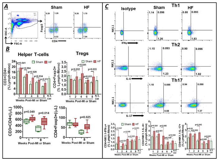
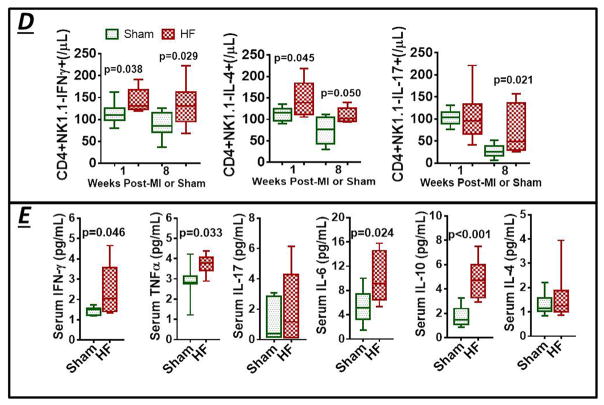
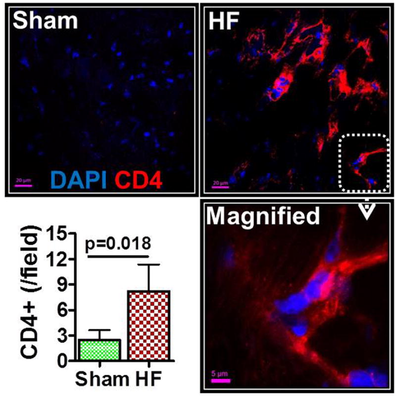
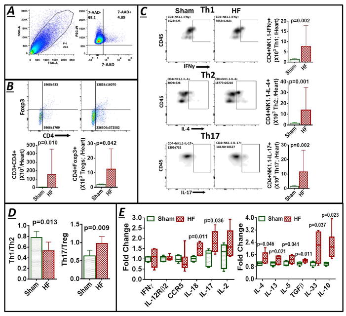
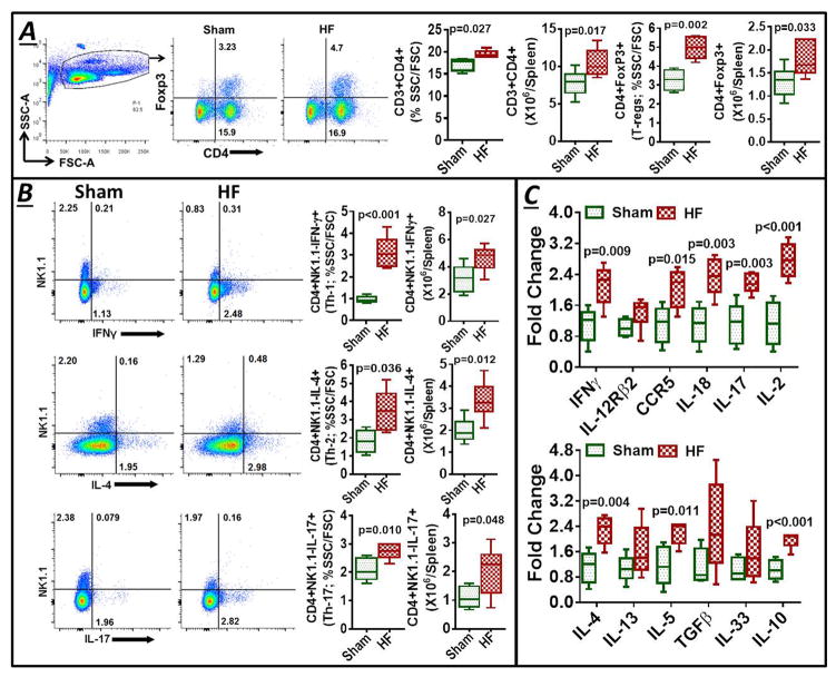
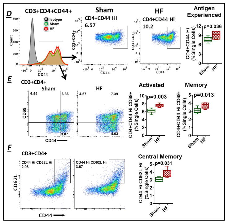
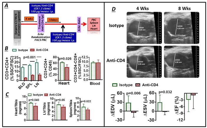
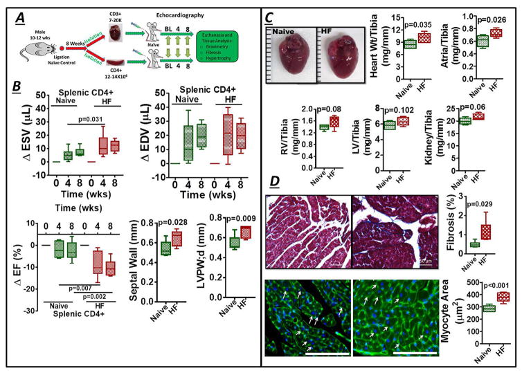
Methods and results: Permanent coronary ligation was performed in adult C57BL/6 mice. When compared with sham-operated mice, mice with HF (8 weeks after ligation) exhibited the following features: (1) significant (P<0.05) expansion of circulating CD3+CD8+ cytotoxic and CD3+CD4+ helper (Th) T lymphocytes, together with increased Th1, Th2, Th17, and regulatory T-cell (Treg) CD4+ subsets; (2) significant expansion of CD8+ and CD4+ T cells in failing myocardium, with increased Th1, Th2, Th17, and Treg CD4+ subsets, marked reduction of the Th1/Th2 ratio, augmentation of the Th17/Treg ratio, and upregulation of Th2 cytokines; and (3) significantly increased Th1, Th2, Th17 cells, and Tregs, in the spleen and mediastinal lymph nodes, with expansion of splenic antigen-experienced effector and memory CD4+ T cells. Antibody-mediated CD4+ T-cell depletion in HF mice (starting 4 weeks after ligation) reduced cardiac infiltration of CD4+ T cells and prevented progressive left ventricular dilatation and hypertrophy, whereas adoptive transfer of splenic CD4+ T cells (and, to a lesser extent, cardiac CD3+ T cells) from donor mice with HF induced long-term left ventricular dysfunction, fibrosis, and hypertrophy in naive recipient mice.

Conclusions: CD4+ T lymphocytes are globally expanded and activated in chronic ischemic HF, with Th2 (versus Th1) and Th17 (versus Treg) predominance in failing hearts, and with expansion of memory T cells in the spleen. Cardiac and splenic T cells in HF are primed to induce cardiac injury and remodeling, and retain this memory on adoptive transfer.

Keywords: T lymphocytes; adaptive immunity; adoptive transfer; heart failure; inflammation.


Figures

Figure 1
Figure 1
(A) Example flow cytometry lymphocyte-monocyte (lymph-mono) SSC/FSC live cell gate and scatter plots for peripheral blood CD3+CD4+ helper and CD3+CD4+Foxp3+ regulatory T-lymphocytes (Tregs) in control and heart failure (HF) mice 8 w after sham-operation or coronary ligation and myocardial infarction (MI). (B) Corresponding group data as %lymphocyte-monocyte gate at serial time points, as well as absolute cell counts (per μL blood) at 1 and 8 w, after operation. (C, D) Representative flow cytometry dot plots and group quantitation of cell frequency (C) and total cell counts (D) for CD4+ Th subsets - Th1 (IFN-γ+), Th2 (IL-4+), and Th17 (IL-17+) - in the same experimental groups. (E) Serum cytokines in mice 8 w after MI (HF) or sham surgery. N.S. Ctrl, non-surgical control.
Figure 1
Figure 1
(A) Example flow cytometry lymphocyte-monocyte (lymph-mono) SSC/FSC live cell gate and scatter plots for peripheral blood CD3+CD4+ helper and CD3+CD4+Foxp3+ regulatory T-lymphocytes (Tregs) in control and heart failure (HF) mice 8 w after sham-operation or coronary ligation and myocardial infarction (MI). (B) Corresponding group data as %lymphocyte-monocyte gate at serial time points, as well as absolute cell counts (per μL blood) at 1 and 8 w, after operation. (C, D) Representative flow cytometry dot plots and group quantitation of cell frequency (C) and total cell counts (D) for CD4+ Th subsets - Th1 (IFN-γ+), Th2 (IL-4+), and Th17 (IL-17+) - in the same experimental groups. (E) Serum cytokines in mice 8 w after MI (HF) or sham surgery. N.S. Ctrl, non-surgical control.
Figure 2
Figure 2
Representative confocal images of CD4 immunostained hearts from sham-operated and HF mice (8 w post-MI). CD4 positivity is indicated by red fluorescence; DAPI-labeled nuclei are blue. A magnified image is shown in the bottom right, and group quantitation of cardiac CD4+ cell abundance in the lower left.
Figure 3
Figure 3
(A) Representative SSC/FSC live cell gate for cardiac mononuclear cells from a failing mouse heart and flow cytometry scatter plots after 7-AAD viability staining (live cells exclude 7-AAD). (B) Example scatter plots, and corresponding group data, for CD4+ helper and CD4+Foxp3+ regulatory T-cell counts in sham and HF hearts. (C) Representative density plots and quantitative group data for Th1, Th17, and Th2 CD4+ T-cell subsets, and (D) Th1/Th2 and Th17/Treg ratios in sham and failing hearts (n = 6–10 per group). (E) Th1- and Th2-related gene expression in border/remote zone myocardium from sham and HF mice (n = 5–6 per group). Fold changes are depicted using β-Actin as the endogenous control.
Figure 4
Figure 4
(A) Example SSC/FSC live cell gate and scatter plots for CD3+CD4+ and CD3+CD4+Foxp3+ Tregs in mononuclear splenocytes isolated from sham and HF mice, and quantitative group data for the same, cell frequency previously published in Ismahil et al. (B) Flow cytometry scatter plots and frequency and cell count group data for CD4+ T-cell subsets in the spleen. (C) Th1 and Th2 related gene expression in splenocytes harvested from sham and HF mice (n=5 per group). Fold changes are shown using 18S rRNA as the endogenous control. (D) Representative flow histograms for splenic CD3+CD4+CD44+ T-cells (Left), scatter plots for identification of antigen-experienced CD44hi cells in sham-operated and HF mice (Middle), and corresponding quantitative group data (Right). (E) Flow cytometry scatter plots and group data for CD4+CD44hiCD69+ activated (effector) T-cells and CD4+CD44hiCD69 total memory T-cells, and (F) CD4+CD44hiCD62Lhi central memory T-cells in splenocytes from sham-operated and HF mice (n = 5–6/group).
Figure 4
Figure 4
(A) Example SSC/FSC live cell gate and scatter plots for CD3+CD4+ and CD3+CD4+Foxp3+ Tregs in mononuclear splenocytes isolated from sham and HF mice, and quantitative group data for the same, cell frequency previously published in Ismahil et al. (B) Flow cytometry scatter plots and frequency and cell count group data for CD4+ T-cell subsets in the spleen. (C) Th1 and Th2 related gene expression in splenocytes harvested from sham and HF mice (n=5 per group). Fold changes are shown using 18S rRNA as the endogenous control. (D) Representative flow histograms for splenic CD3+CD4+CD44+ T-cells (Left), scatter plots for identification of antigen-experienced CD44hi cells in sham-operated and HF mice (Middle), and corresponding quantitative group data (Right). (E) Flow cytometry scatter plots and group data for CD4+CD44hiCD69+ activated (effector) T-cells and CD4+CD44hiCD69 total memory T-cells, and (F) CD4+CD44hiCD62Lhi central memory T-cells in splenocytes from sham-operated and HF mice (n = 5–6/group).
Figure 5
Figure 5
(A) Protocol used for antibody-mediated CD4+ T-cell depletion in HF mice; PBC, peripheral blood cells. (B) Quantitative group data for CD4+ T-cells in blood, spleen, mediastinal LNs and hearts, and blood CD8+ T-cells, in HF mice treated with either anti-CD4 antibody or isotype control. (C) normalized heart, LV and spleen weight in HF mice treated as in B. (D) Representative end-diastolic echocardiographic images from HF mice before (4 w post-MI) and after (8 w post-MI) treatment with either isotype or anti-CD4 antibody, and quantitative group data for changes in LV end-diastolic and end-systolic volume (EDV and ESV) and ejection fraction (EF) (n = 6–10/group).
Figure 6
Figure 6
(A) Protocol for adoptive transfer of splenic CD4+ and cardiac CD3+ T-cells from naïve control and HF mice to normal recipients. (B) Changes in LV ESV, EDV, and EF in recipient mice 4 and 8 w after splenic CD4+ T-cell transfer, along with measured septal and posterior wall (PW) thickness at 8 w. (C) Gross images of hearts isolated from mice 8 w after receiving either control or HF-activated splenic CD4+ T-cells and normalized gravimetric data for the tissues indicated; RV, right ventricle. (D) Representative Masson’s trichrome stains and quantitation of fibrosis (Top), and wheat germ-agglutinin stains (cell membranes are green, nuclei are blue with DAPI counterstaining; scale bar 200 μm) and quantitative data for myocyte size (Bottom), from mouse hearts 8 w after transfer of either naïve control or HF-activated splenic CD4+ T-cells. Arrows indicate example myocytes used for determination of cross-sectional area.

References

    1. Mann DL. Inflammatory mediators and the failing heart: Past, present, and the foreseeable future. Circ Res. 2002;91:988–998. - PubMed
    1. Prabhu SD, Chandrasekar B, Murray DR, Freeman GL. Beta-adrenergic blockade in developing heart failure: Effects on myocardial inflammatory cytokines, nitric oxide, and remodeling. Circulation. 2000;101:2103–2109. - PubMed
    1. Chung ES, Packer M, Lo KH, Fasanmade AA, Willerson JT. Randomized, double-blind, placebo-controlled, pilot trial of infliximab, a chimeric monoclonal antibody to tumor necrosis factor-α, in patients with moderate-to-severe heart failure: Results of the anti-TNF therapy against congestive heart failure (ATTACH) trial. Circulation. 2003;107:3133–3140. - PubMed
    1. Mann DL. Innate immunity and the failing heart: The cytokine hypothesis revisited. Circ Res. 2015;116:1254–1268. - PMC - PubMed
    1. Mann DL, McMurray JJ, Packer M, Swedberg K, Borer JS, Colucci WS, Djian J, Drexler H, Feldman A, Kober L, Krum H, Liu P, Nieminen M, Tavazzi L, van Veldhuisen DJ, Waldenstrom A, Warren M, Westheim A, Zannad F, Fleming T. Targeted anticytokine therapy in patients with chronic heart failure: Results of the randomized etanercept worldwide evaluation (renewal) Circulation. 2004;109:1594–1602. - PubMed

MeSH terms