Review
. 2017 Jun 1;7(6):a025486.
doi: 10.1101/cshperspect.a025486.

Malaria Parasite Liver Infection and Exoerythrocytic Biology

Affiliations
Review

Malaria Parasite Liver Infection and Exoerythrocytic Biology

Ashley M Vaughan et al. Cold Spring Harb Perspect Med. .

Abstract

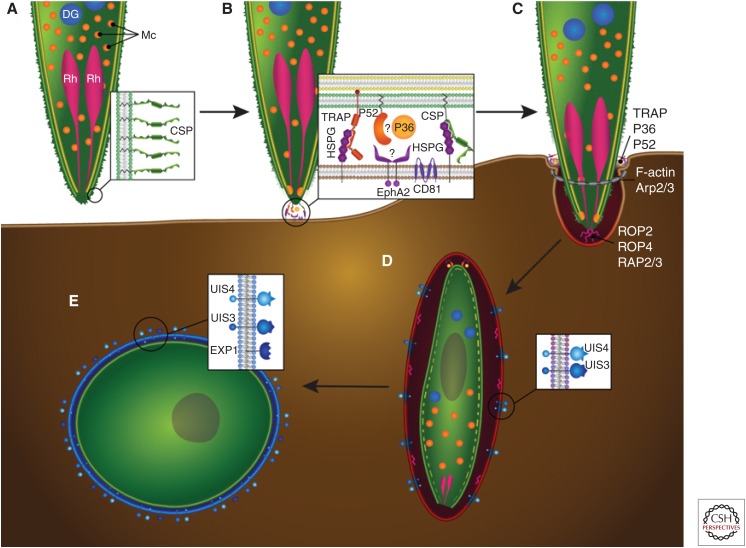
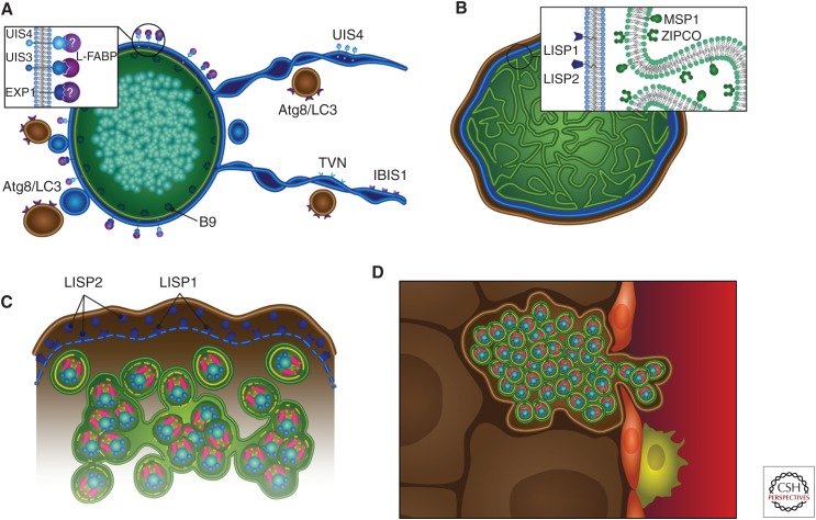
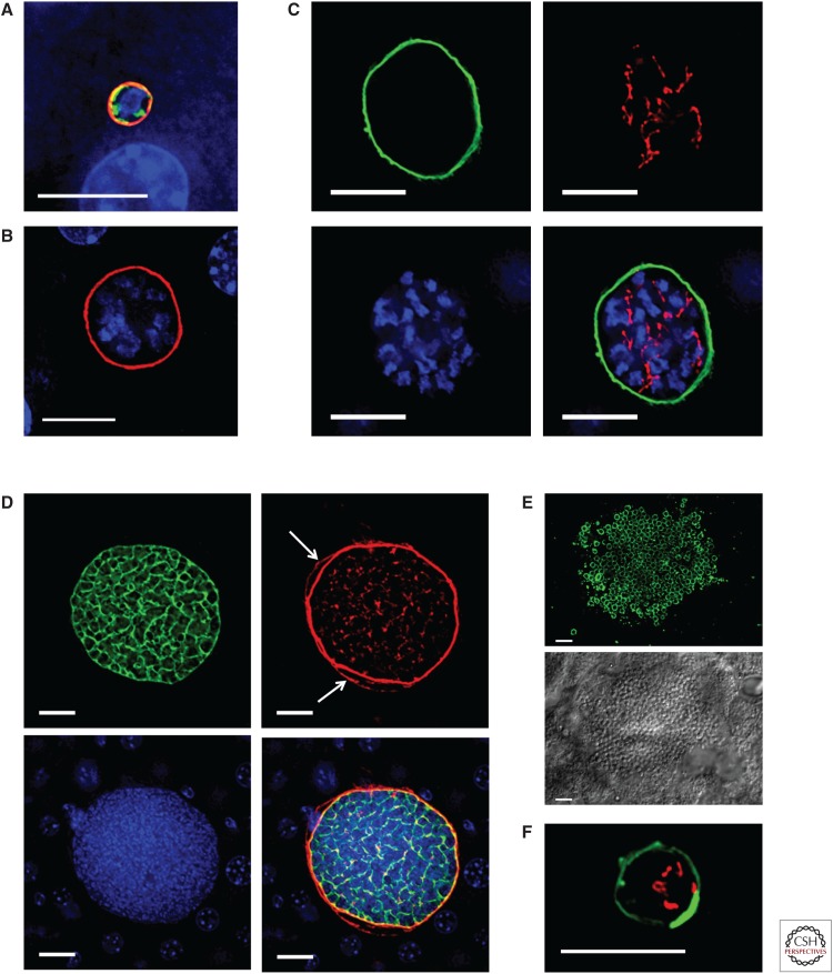
In their infection cycle, malaria parasites undergo replication and population expansions within the vertebrate host and the mosquito vector. Host infection initiates with sporozoite invasion of hepatocytes, followed by a dramatic parasite amplification event during liver stage parasite growth and replication within hepatocytes. Each liver stage forms up to 90,000 exoerythrocytic merozoites, which are in turn capable of initiating a blood stage infection. Liver stages not only exploit host hepatocyte resources for nutritional needs but also endeavor to prevent hepatocyte cell death and detection by the host's immune system. Research over the past decade has identified numerous parasite factors that play a critical role during liver infection and has started to delineate a complex web of parasite-host interactions that sustain successful parasite colonization of the mammalian host. Targeting the parasites' obligatory infection of the liver as a gateway to the blood, with drugs and vaccines, constitutes the most effective strategy for malaria eradication, as it would prevent clinical disease and onward transmission of the parasite.


Figures

Figure 1.
Figure 1.
Sporozoite invasion of host hepatocytes. Graphic representation of the invasion steps that culminate in the productive infection of the hepatocyte host cell. (A) The surface of the motile sporozoite is coated with circumsporozoite protein (CSP) (shown in inset), which engages heparin sulfate proteoglycans (HSPGs) on the hepatocyte surface. The apical end of the sporozoite contains organelles whose contents are essential for hepatocyte invasion: rhoptries (Rh, pink), micronemes (Mc, orange), and likely dense granules (DG, blue), although the latter have not been unequivocally identified. (B,C) Contact with an invasion-permissive hepatocyte (brown) triggers CSP processing, which, via an unknown mechanism, triggers the release of invasion-essential proteins from the micronemes (inset). These proteins include thrombospondin-related adhesive protein (TRAP), which binds to HSPGs on the hepatocyte cell surface and to the sporozoite internal glideosome complex (not shown) via its cytoplasmic domain to provide traction for the invading sporozoite at the moving junction (also known as the tight junction, shown as a gray ring). The microneme proteins P52 and P36 are also involved in the invasion process and may interact with each other as well as interacting with the hepatocyte Ephrin A2 receptor (EphA2). The P52/P36/EphA2 axis appears to be critical for the formation of the parasitophorous vacuole (PV). The hepatocyte receptor CD81 is also important for the invasion process and PV formation, but it is not clear whether the sporozoite directly interacts with it. The inner membrane complex (IMC) (in yellow) anchors the internal glideosome complex, allowing for sporozoite movement into the hepatocyte. Hepatocyte invasion occurs through the moving junction at the point of entry and is accompanied by the polymerization of host F-actin in association with Arp2/3. Invasion results in the invagination of the hepatocyte plasma membrane and the release of rhoptry proteins, including ROP2, ROP4, and RAP2/3. (D) Successful invasion results in the sporozoite residing within a PV surrounded by a parasitophorous vacuole membrane (PVM) of hepatocyte origin. The PVM is extensively modified by the parasite and the putative dense granule proteins UIS3 and UIS4 are released and trafficked to the PVM, as is EXP-1 (inset). During sporozoite dedifferentiation, the IMC (dashed yellow line) and the apical organelles are broken down. (E) The nascent liver stage trophozoite resides within a PV, has its own parasite plasma membrane still coated by CSP (green), and is surrounded by a PVM (blue).
Figure 2.
Figure 2.
Liver stage development. Graphic representation of liver stage maturation that leads to the formation of exoerythrocytic merozoites. (A) The tubulovesicular network (TVN) (blue)—membrane-bound extensions and whorls that emanate from the parasitophorous vacuole membrane (PVM)—interacts with the hepatocyte’s autophagosomes (which express Atg8/LC3) likely for nutrient uptake. Parasite proteins expressed on the PVM/TVN include IBIS1, EXP1, UIS4, and UIS3, which has been shown to interact with host L-FABP. The parasite plasma membrane (PPM)-associated protein B9 is known to be important for liver stage development. (B) As the liver stage parasite matures, multiple invaginations of the PPM occur (cytomere formation), and this is accompanied by the expression of MSP1 and ZIPCO. Additionally, LISP1 and LISP2 expression occur on the PVM. (C) Toward the end of liver stage development, individual exoerythrocytic merozoites begin to form, the PVM breaks down (a process that relies on LISP1), and LISP2 is released into the host hepatocyte. (D) Merosomes, merozoites surrounded by hepatocyte plasma membrane, are released into the bloodstream through the liver sinusoid, which is demarcated by epithelial cells (red) and liver resident macrophages, the Kupffer cells (yellow).
Figure 3.
Figure 3.
Immunofluorescence images of liver stage development. (AE) Plasmodium yoelii liver stage development in a mouse liver. (A) The spherical, early liver stage parasite, 12 h after sporozoite invasion, is significantly smaller than the host hepatocyte nucleus (blue). The liver stage parasitophorous vacuole (PVM) is shown in red (UIS4 expression), the endoplasmic reticulum (ER) is shown in green (BiP expression), and the parasite contains a single nuclear center (blue). (B) By 24 h, the P. yoelii liver stage parasite (the UIS4-positive PVM is shown in red) has entered schizogony (multiple centers of nuclear replication [blue]). (C) At 30 h of development, the P. yoelii liver stage (the UIS4 PVM is shown in green) has undergone multiple rounds of nuclear replication (blue) and contains a highly branched apicoplast (acyl carrier protein expression, shown in red). (D) Late in P. yoelii liver stage development (48 h), the parasite plasma membrane (PPM) undergoes extensive invagination resulting in cytomere formation (MSP1, green). LISP2 (red) is expressed on the PVM as well as the hepatocyte membrane (white arrows). The nuclei (blue) of the individual parasites can be seen within the liver stage parasite. (E) At the end of liver stage development (52 h), individual exoerythrocytic merozoites (MSP1 expression in green, upper panel, delineates the merozoite membrane) can be seen within the mature liver stage parasite (differential interference contrast, lower panel). (F) Image of a P. vivax hypnozoite in a humanized mouse liver. The hypnozoite remains latent, is small, and does not replicate its DNA, and its PVM has a unique UIS4-positive prominence (green). The hypnozoite mitochondrion (HSP60 expression) is shown in red and is branched. Scale bars, 10 µm.

References

    1. Albuquerque SS, Carret C, Grosso AR, Tarun AS, Peng X, Kappe SH, Prudencio M, Mota MM. 2009. Host cell transcriptional profiling during malaria liver stage infection reveals a coordinated and sequential set of biological events. BMC Genomics 10: 270. - PMC - PubMed
    1. Aly AS, Mikolajczak SA, Rivera HS, Camargo N, Jacobs-Lorena V, Labaied M, Coppens I, Kappe SH. 2008. Targeted deletion of SAP1 abolishes the expression of infectivity factors necessary for successful malaria parasite liver infection. Mol Microbiol 69: 152–163. - PMC - PubMed
    1. Annoura T, van Schaijk BC, Ploemen IH, Sajid M, Lin JW, Vos MW, Dinmohamed AG, Inaoka DK, Rijpma SR, van Gemert GJ, et al. 2014. Two Plasmodium 6-Cys family-related proteins have distinct and critical roles in liver-stage development. FASEB J 28: 2158–2170. - PubMed
    1. Azuma H, Paulk N, Ranade A, Dorrell C, Al-Dhalimy M, Ellis E, Strom S, Kay MA, Finegold M, Grompe M. 2007. Robust expansion of human hepatocytes in Fah-/-/Rag2-/-/Il2rg-/- mice. Nat Biotechnol 25: 903–910. - PMC - PubMed
    1. Baer K, Klotz C, Kappe SH, Schnieder T, Frevert U. 2007. Release of hepatic Plasmodium yoelii merozoites into the pulmonary microvasculature. PLoS Pathog 3: e171. - PMC - PubMed