. 2017 Feb;5(4):e13154.
doi: 10.14814/phy2.13154. Epub 2017 Feb 27.

Effects of functional decoupling of a leg in a model of stick insect walking incorporating three ipsilateral legs

Affiliations

Effects of functional decoupling of a leg in a model of stick insect walking incorporating three ipsilateral legs

Tibor I Tóth et al. Physiol Rep. 2017 Feb.

Abstract

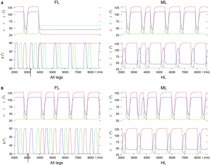
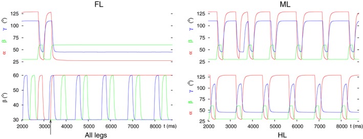
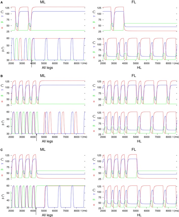
Legged locomotion is a fundamental form of activity of insects during which the legs perform coordinated movements. Sensory signals conveying position, velocity and load of a leg are sent between the thoracic ganglia where the local control networks of the leg muscles are situated. They affect the actual state of the local control networks, hence the stepping of the legs. Sensory coordination in stepping has been intensively studied but important details of its neuronal mechanisms are still unclear. One possibility to tackle this problem is to study what happens to the coordination if a leg is, reversibly or irreversibly, deprived of its normal function. There are numerous behavioral studies on this topic but they could not fully uncover the underlying neuronal mechanisms. Another promising approach to make further progress here can be the use of appropriate models that help elucidate those coordinating mechanisms. We constructed a model of three ipsilateral legs of a stick insect that can mimic coordinated stepping of these legs. We used this model to investigate the possible effects of decoupling a leg. We found that decoupling of the front or the hind leg did not disrupt the coordinated walking of the two remaining legs. However, decoupling of the middle leg yielded mixed results. Both disruption and continuation of coordinated stepping of the front and hind leg occurred. These results agree with the majority of corresponding experimental findings. The model suggests a number of possible mechanisms of decoupling that might bring about the changes.

Keywords: Insect locomotion; network model; neuromuscular control.


Figures

Figure 1
Figure 1
(A) Schematic illustration of a (middle) leg of the stick insect showing three antagonistic muscle pairs that are the most important ones for walking. (Adapted with permission from M.Gruhn unpublished observations.) (B) Lifted, protracted, and extended position of the leg. (C) The leg is on the ground, retracted and flexed. (D) The model of three ipsilateral legs (3‐leg model). Three copies of nearly identical neuro‐muscular networks sitting in the three thoracic segments: the pro‐, meso‐, and metathoracic one, as indicated. They correspond to the front leg (FL), middle leg (ML), and hind leg (HL), respectively. Local networks at one thoracic segment: PR (protractor‐retractor), LD (levator‐depressor), and EF (extensor‐flexor), as indicated, controlling the activity of three antagonistic muscle pairs: m. protractor and retractor coxae (Pro. m. and Ret. m.); m. levator and depressor trochanteris (Lev. m. and Dep. m.); and m. extensor and flexor tibiae (Ext. m. and Flex. m.). CPG in each network: central pattern generator consisting of a pair of mutually inhibitory nonspiking neurons: C1–C2 in the PR, C3–C4 in the LD, and C5–C6 in the EF control network at the prothoracic segment; The arrangement is the same in the two other thoracic segments. g app,1, g app,2 etc.: (central) input to the CPG neurons (individually variable). MN1P, MN2R etc.: motoneurons driving the corresponding muscles; gMN: uniform excitatory input to all motoneurons. IN1‐IN2 etc.: premotor interneurons; g d1, g d2 etc.: (individually variable) inhibitory inputs to these interneurons. IN3‐IN4 etc.: interneurons conveying intrasegmental sensory signals to the corresponding CPG neurons from the other local networks. Hexagons with β or γ in them: sources of sensory signals encoding position, load, and ground contact. Pentagons with dβ in them: sources of sensory signals encoding (angular) velocity. The arrows originating at them identify the synaptic pathways they affect. (For a detailed explanation see Tóth and Daun‐Gruhn 2016). Other symbols: empty triangles: excitatory synapses; filled circles: inhibitory synapses. Arrows from muscles to the hexagons symbolize that the sensory signals arise because of mechanical movement due to muscle activity. Intersegmental thick lines connecting the LD systems: inhibitory synaptic pathways from the posterior segment to the next anterior one. g inh3, g inh9: actual synaptic strengths exerting influence on the CPG on which they converge (CPG C3‐C4 and CPG C9‐C10, respectively). These synaptic strengths are completely determined by the actual values of β in the next posterior segment. Note that there is no such inhibitory synaptic connection on the LD CPG of the metathoracic (HL) segment.
Figure 2
Figure 2
The three basic ways of decoupling one leg from the coordination mechanism of the three legs in the model, exemplified by decoupling the front leg. The numbers 1, 2, and 3 denote these possibilities. 1: decoupling at the intersegmental coordinating synapses from the hind and middle leg. 2: decoupling by changing the (central) drive to the CPG neurons of the LD local network, that is, changing the value of the corresponding conductances gapp. 3: decoupling by changing the input (conductances g d5, g d6) to the premotor INs in the LD local network. ML, middle leg; HL, hind leg. Other notations are the same as in Figure 1D.
Figure 3
Figure 3
Angular movements of the three legs before and after decoupling of the front leg by perturbing the intersegmental coordination. In all (A, B) and (C) panel FL: angular movements of the front leg as time functions (α(t), β(t), γ(t)) defined in Methods); the ranges for these angles are: α: 28° (maximal protraction) – 128° (maximal retraction), β: 30° (on the ground) – 60° (maximal elevation), γ: 45° (maximal extension) – 110° (maximal flexion); panel all legs: vertical movement of the femur (β angles) of the front (red curve), the middle (green curve) and the hind (blue curve) leg; black arrow: start of the decoupling of the front leg; panel ML: angular movements of the middle leg as time functions (in analogy to panel FL), and panel HL: angular movements of the hind leg as time functions (in analogy to panel FL). The two latter panels show the state of the intraleg coordination in the unaffected legs. This also holds for the panels on the right‐hand side in the subsequent figures. (A) Tetrapod and tripod coordination patterns and the transition between them in the 3‐leg model (control case). (B) Decoupling by changing the intersegmental synapse permanently to an excitatory one. (C) Decoupling FL by switching off the intersegmental synapse (setting its conductance permanently to zero). Note the different steady‐state position of the front leg in B (lifted, protracted, and extended, cf. Fig. 1B) and C (on the ground, retracted and flexed, cf. Fig. 1C). Note also the coordinated (alternating) stepping of the middle and the hind leg in these panels after decoupling of the front leg.
Figure 4
Figure 4
Successful and failed decoupling of the front leg by changing the (central) synaptic drives to the CPG of the LD system of the front leg. Depending on the phase of the stepping period at which the decoupling command is evoked, the decoupling may succeed or fail. (A) successful decoupling. Note the steady‐state position of the front leg. (B) failed decoupling. The same permanent change to the drives (g app3 and g app4) as in A remains ineffective. All notations are the same as in Figure 3.
Figure 5
Figure 5
Decoupling of the front leg by inhibiting the inhibitory IN to the levator MN and fully disinhibiting the inhibitory IN to the depressor MN. Note that these changes already sufficed to bring the front leg in the ‘desired’ spatial position: lifted, protracted, stretched. The coordinated stepping of the hind and middle leg continues. All notations are the same as in Figure 3.
Figure 6
Figure 6
Decoupling of the hind leg by setting the input to both LD CPG neurons to zero. Two types of results emerged: (A) Hind leg on the ground, retracted, and stretched. (B) Hind leg lifted, protracted, flexed. In both cases, the coordinated stepping of the front and middle leg continued. All notations are the same as in Figure 3.
Figure 7
Figure 7
Decoupling of the hind leg by inhibiting the inhibitory IN to the de‐pressor MN and disinhibiting the inhibitory IN to the levator MN. (A) Starting with tripod coordination pattern. (B) Starting with tetrapod coordination pattern. Note that the hind leg behaves the same way in both cases attaining the spatial position seen in the experiments. The coordinated stepping of the front and middle leg continues. All notations are the same as in Figure 3.
Figure 8
Figure 8
Decoupling of the middle leg at the intersegmental synapse by setting it (with conductance g inh9) permanently excitatory. (A) Starting with tripod coordination pattern. As it can be seen, the middle leg does not stop its rhythmic stepping but that occurs in synchrony with the hind leg's. In addition, the intervals in which both the front leg and the middle leg are lifted partly overlap. An “unusual” coordination pattern is produced. (B) Starting with tetrapod coordination pattern. Here, after the decoupling command (arrow), the middle leg exerts slow rhythmic stepping, the front leg, however, stays permanently lifted, protracted, and stretched. The hind leg continues its normal stepping. In this case, too, the coordination pattern is “unusual”. All notations are the same as in Figure 3.
Figure 9
Figure 9
Decoupling of the middle leg by permanently changing the input to its LD CPG. Three typical results are illustrated. The first result was obtained by applying (strong) excitatory input to the levator and (strong) inhibitory input to the depressor CPG neuron. The second and third one arose, when there was no input to the CPG (both input conductances were set to zero; Figure 1D). (A) The middle leg does not have proper ground contact; the front leg remains lifted, protracted, and stretched; the hind leg continues its normal stepping. This result was obtained with both starting coordination patterns (tripod, tetrapod). The result displayed is with the starting coordination pattern tripod. (B) All three legs continue stepping but the front and middle leg step in synchrony. This result could only be seen with starting tripod coordination pattern. (C) The middle and the hind leg continue tetrapod stepping but the front leg remains permanently lifted (protracted and stretched). This result clearly shows a failed decoupling of the middle leg but a successful decoupling of the front leg. It occurred only with starting tetrapod coordination pattern. All notations are the same as in Figure 3.
Figure 10
Figure 10
Decoupling of the middle leg via premotor inhibitory INs. (A) Premotor IN to the depressor MN is inhibited, while the one to the levator MN is disinhibited. As a result, the middle leg permanently remains on the ground (retracted, flexed). The front leg stays lifted (protracted and stretched), while the hind leg continues normal stepping. (B) The inputs to the premotor INs are the same as in A but the decoupling command arrives at a different phase of the stepping period. As a result, the front leg and the hind leg now perform coordinated stepping. (C) Premotor IN to the depressor MN is disinhibited, while the one to the levator MN is inhibited. The middle leg therefore stays lifted (protracted and stretched). The front leg also stays lifted as in A. Here, the starting coordination pattern in A was tetrapod, while in B and C tripod. All notations are the same as in Figure 3.

Similar articles

Cited by

References

    1. Bässler, U. 1977. Sensory control of leg movement in the stick insect Carausius morosus. Biol. Cybern. 25:61–72. - PubMed
    1. Bässler, U . 1983. Neural basis of elementary behavior in stick insects. Springer Verlag, Berlin‐ Heidelberg, Germany.
    1. Bässler, U . 1993. The walking‐ (and searching‐) pattern generator of stick insects, a modular system composed of reflex chains and endogenous oscillators. Biol. Cybern. 69:305–317.
    1. Berg, E. , Büschges A., and Schmidt J.. 2011. Single perturbations cause sustained changes in searching behavior in stick insects. J. Exp. Biol. 216:1064–1074. - PubMed
    1. Borgmann, A. , Scharstein H., and Buschges A.. 2007. Intersegmental coordination: influence of a single walking leg on the neighboring segments in the stick insect walking system. J. Neurophysiol. 98:1685–1696. - PubMed

LinkOut - more resources