. 2017 Feb 10:12:1183-1200.
doi: 10.2147/IJN.S126887. eCollection 2017.

Heterogeneous dimer peptide-conjugated polylysine dendrimer-Fe3O4 composite as a novel nanoscale molecular probe for early diagnosis and therapy in hepatocellular carcinoma

Affiliations

Heterogeneous dimer peptide-conjugated polylysine dendrimer-Fe3O4 composite as a novel nanoscale molecular probe for early diagnosis and therapy in hepatocellular carcinoma

Jian-Min Shen et al. Int J Nanomedicine. .

Abstract

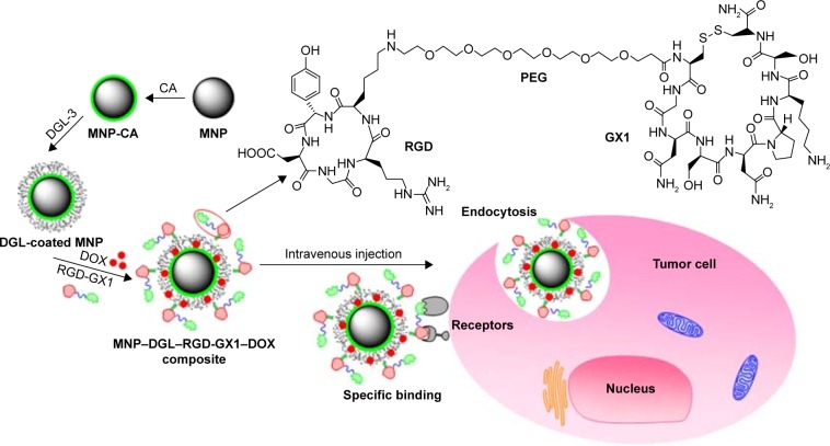
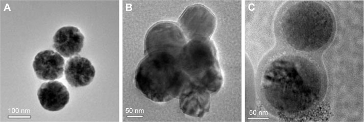
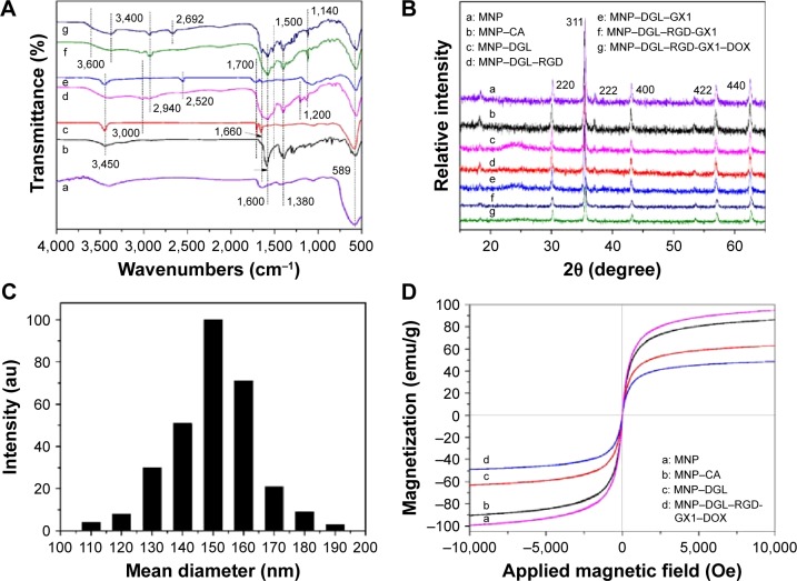
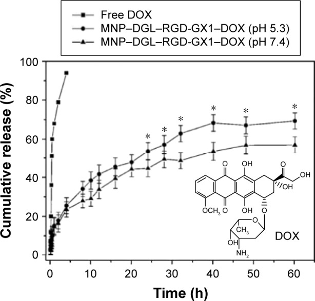
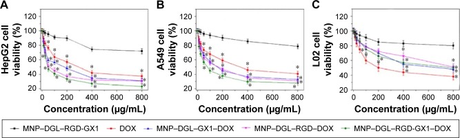
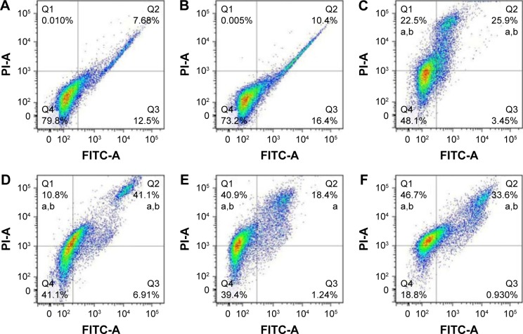
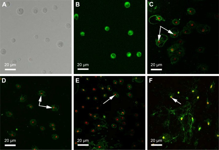
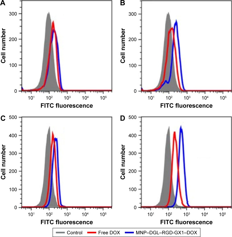
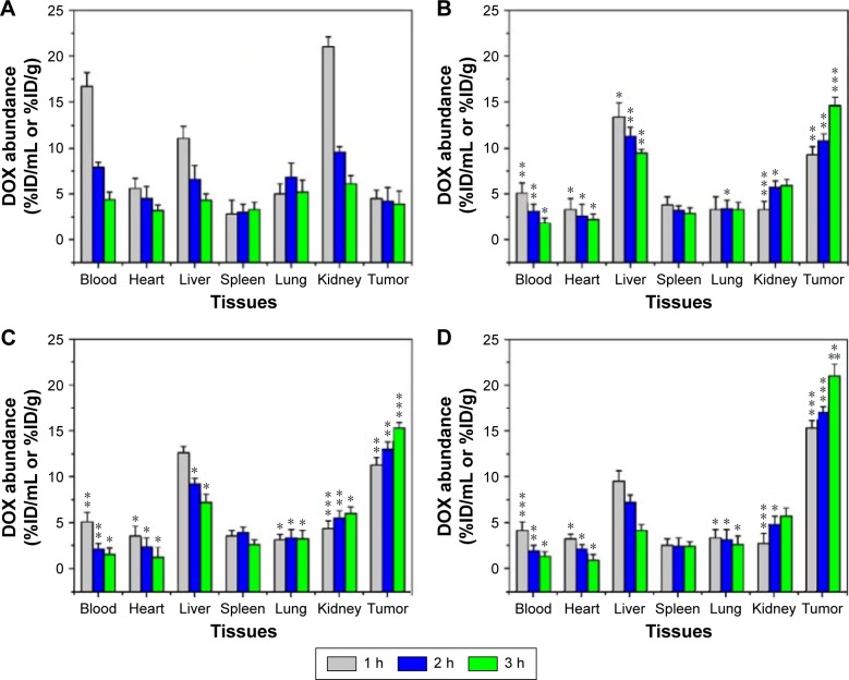
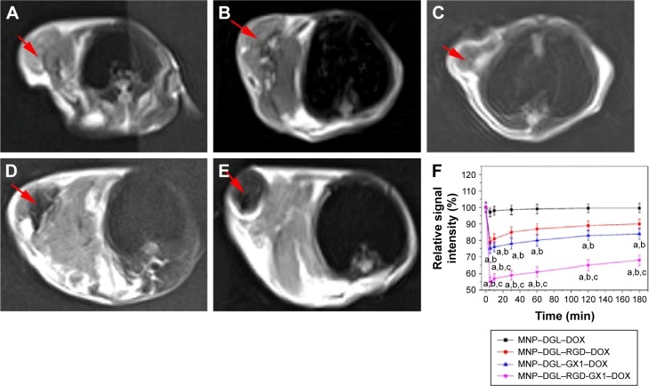
A novel nanoscale molecular probe is formulated in order to reduce toxicity and side effects of antitumor drug doxorubicin (DOX) in normal tissues and to enhance the detection sensitivity during early imaging diagnosis. The mechanism involves a specific targeting of Arg-Gly-Asp peptide (RGD)-GX1 heterogeneous dimer peptide-conjugated dendrigraft poly-l-lysine (DGL)-magnetic nanoparticle (MNP) composite by αvβ3-integrin/vasculature endothelium receptor-mediated synergetic effect. The physicochemical properties of the nanoprobe were characterized by using transmission electron microscope, Fourier transform infrared spectroscopy, X-ray diffraction, dynamic light scattering (DLS), and vibrating sample magnetometer. The average diameter of the resulting MNP-DGL-RGD-GX1-DOX nanoparticles (NPs) was ~150-160 nm by DLS under simulate physiological medium. In the present experimental system, the loading amount of DOX on NPs accounted for 414.4 mg/g for MNP-DGL-RGD-GX1-DOX. The results of cytotoxicity, flow cytometry, and cellular uptake consistently indicated that the MNP-DGL-RGD-GX1-DOX NPs were inclined to target HepG2 cells in selected three kinds of cells. In vitro exploration of molecular mechanism revealed that cell apoptosis was associated with the overexpression of Fas protein and the significant activation of caspase-3. In vivo magnetic resonance imaging and biodistribution study showed that the MNP-DGL-RGD-GX1-DOX formulation had high affinity to the tumor tissue, leading to more aggregation of NPs in the tumor. In vivo antitumor efficacy research verified that MNP-DGL-RGD-GX1-DOX NPs possessed significant antitumor activity and the tumor inhibitory rate reached 78.5%. These results suggested that NPs could be promising in application to early diagnosis and therapy in hepatocellular carcinoma as a specific nanoprobe.

Keywords: hepatocellular carcinoma (HCC); heterogeneous dimer peptide (HDP); magnetic nanoparticles (MNPs); molecular probe; targeting.


Conflict of interest statement

Disclosure The authors report no conflicts of interest in this work.

Figures

Figure 1
Figure 1
Schematic diagram of designing heterogeneous dimer peptide-conjugated polylysine dendrimer-Fe3O4 nanoparticles, targeting toward hepatocellular carcinoma. Abbreviations: DGL, dendrigraft poly-l-lysine; MNP, magnetic nanoparticle; CA, citric acid; DOX, doxorubicin; RGD, Arg-Gly-Asp peptide; GX1, cyclo[-Cys-Gly-Asn-Ser-Asn-Pro-Lys-Ser-Cys] peptide.
Figure 2
Figure 2
TEM images of (A) MNP–CA, (B) MNP–DGL, and (C) MNP–DGL–RGD-GX1–DOX composite. Abbreviations: MNP, magnetic nanoparticle; CA, citric acid; DGL, dendrigraft poly-l-lysine; RGD, Arg-Gly-Asp peptide; GX1, cyclo[-Cys-Gly-Asn-Ser-Asn-Pro-Lys-Ser-Cys] peptide; DOX, doxorubicin.
Figure 3
Figure 3
(A) Fourier transform infrared spectroscopy spectra of MNP (a), MNP-CA (b), MNP-DGL (c), MNP-DGL-RGD (d), MNP-DGL-GX1 (e), MNP-DGL-RGD-GX1 (f), and MNP-DGL-RGD-GX1-DOX (g) NPs. (B) XRD patterns. (C) The size distribution of the MNP–DGL–RGD-GX1–DOX measured by DLS. (D) VSM magnetization curves. Abbreviations: MNP, magnetic nanoparticle; CA, citric acid; DGL, dendrigraft poly-l-lysine; RGD, Arg-Gly-Asp peptide; GX1, cyclo[-Cys-Gly-Asn-Ser-Asn-Pro-Lys-Ser-Cys] peptide; DOX, doxorubicin; NP, nanoparticle; XRD, X-ray diffraction; DLS, dynamic light scattering; VSM, vibrating sample magnetometer.
Figure 4
Figure 4
Release profiles of DOX from MNP–DGL–RGD-GX1–DOX NPs in a simulated normal body fluid (pH 7.4, 50 mM PBS containing 0.6 mM HSA and 0.1 M NaCl) and an acidic solution (pH 5.3 50 mM PBS containing 0.6 mM HSA and 0.1 M NaCl) at 37°C±1°C. Free DOX curve was conducted to testify that DOX molecules were not trapped inside dialysis bag. The inset in the figure indicates the molecular structure of DOX. The data are expressed as mean ± SD (n=3). *P<0.05 versus pH 7.4 group according to ANOVA. Abbreviations: MNP, magnetic nanoparticle; DGL, dendrigraft poly-l-lysine; RGD, Arg-Gly-Asp peptide; GX1, cyclo[-Cys-Gly-Asn-Ser-Asn-Pro-Lys-Ser-Cys] peptide; DOX, doxorubicin; NP, nanoparticle; PBS, phosphate buffer solution; HAS, human serum albumin; SD, standard deviation; ANOVA, analysis of variance.
Figure 5
Figure 5
Relative cell viabilities of (A) HepG2 cells, (B) A549 cells, and (C) L02 cells incubated with different concentrations of empty NPs and DOX-loaded NPs for 48 h, respectively. The concentration of free DOX was determined according to the contents loaded in NPs and their release efficiencies. The data are expressed as mean ± SD (n=3). *P<0.05 versus MNP–DGL–RGD-GX1 group according to ANOVA. Abbreviations: NP, nanoparticle; DOX, doxorubicin; SD, standard deviation; MNP, magnetic nanoparticle; DGL, dendrigraft poly-l-lysine; RGD, Arg-Gly-Asp peptide; GX1, cyclo[-Cys-Gly-Asn-Ser-Asn-Pro-Lys-Ser-Cys] peptide; ANOVA, analysis of variance.
Figure 6
Figure 6
Apoptosis analysis of representative HepG2 cells incubated with the different NPs (400 μg/mL) or free DOX solutions (3 μg/mL) corresponding to the DOX concentrations loaded on NPs for 48 h. (A) Control HepG2 cells (untreated with NPs or DOX), (B) HepG2 cells + MNP–DGL–RGD-GX1, (C) HepG2 cells + free DOX, (D) HepG2 cells + MNP–DGL–GX1–DOX, (E) HepG2 cells + MNP–DGL–RGD–DOX, (F) HepG2 cells + MNP–DGL–RGD-GX1–DOX. aP<0.05 versus control and bP<0.05 versus MNP–DGL–RGD-GX1 group according to ANOVA. Abbreviations: NP, nanoparticle; DOX, doxorubicin; MNP, magnetic nanoparticle; DGL, dendrigraft poly-l-lysine; RGD, Arg-Gly-Asp peptide; GX1, cyclo[-Cys-Gly-Asn-Ser-Asn-Pro-Lys-Ser-Cys] peptide; ANOVA, analysis of variance; PI, propidium iodide.
Figure 7
Figure 7
Laser scanning confocal images of the HepG2 cells incubated with the different NPs (400 μg/mL) or free DOX solutions (3 μg/mL) corresponding to the DOX concentrations loaded on NPs for 48 h. (A) Control HepG2 cells, (B) HepG2 cells + MNP–DGL–RGD-GX1, (C) HepG2 cells + free DOX, (D) HepG2 cells + MNP–DGL–GX1–DOX, (E) HepG2 cells + MNP–DGL–RGD–DOX, (F) HepG2 cells + MNP–DGL–RGD-GX1–DOX. (BF) The cytoplasms exist green fluorescence, (CF) The nuclei have red fluorescence. The white arrows are cell nucleus with red fluorescence. Excitation laser wavelength =488 nm. Abbreviations: NP, nanoparticle; DOX, doxorubicin; MNP, magnetic nanoparticle; DGL, dendrigraft poly-l-lysine; RGD, Arg-Gly-Asp peptide; GX1, cyclo[-Cys-Gly-Asn-Ser-Asn-Pro-Lys-Ser-Cys] peptide; ANOVA, analysis of variance.
Figure 8
Figure 8
(A and B) The expression of Fas protein and (C and D) the activities of caspase-3 in HepG2 cells treated with MNP–DGL–RGD-GX1–DOX NPs and free DOX solutions for 48 h, respectively. (A and C) Illustrate the HepG2 cells treated with NPs and free DOX solutions (0.16 μg/mL) corresponding to the DOX concentrations loaded on NPs, respectively. (C and D) Illustrate the HepG2 cells treated with NPs and free DOX solutions (1.25 μg/mL) corresponding to the DOX concentrations loaded on NPs, respectively. Abbreviations: MNP, magnetic nanoparticle; DGL, dendrigraft poly-l-lysine; RGD, Arg-Gly-Asp peptide; GX1, cyclo[-Cys-Gly-Asn-Ser-Asn-Pro-Lys-Ser-Cys] peptide; DOX, doxorubicin; NP, nanoparticle.
Figure 9
Figure 9
DOX abundance in percent injected dose per milliliter (%ID/mL) of blood or per gram (%ID/g) of tissue from the male HepG2-bearing Balb/c mice at defined time periods (1, 2, and 3 h) post-intravenous injection of (A) free DOX corresponding to the DOX concentration loaded on NPs in sterile PBS, (B) MNP–DGL–RGD–DOX, (C) MNP–DGL–GX1–DOX, and (D) MNP–DGL–RGD-GX1–DOX NPs at a dose of 2 mg/kg body weight. The data are expressed as mean ± SD (n=5 mice at each time point, each group). *P<0.05, **P<0.01, and ***P<0.001 versus free DOX group according to ANOVA. Abbreviations: DOX, doxorubicin; NP, nanoparticle; PBS, phosphate buffer solution; MNP, magnetic nanoparticle; DGL, dendrigraft poly-l-lysine; RGD, Arg-Gly-Asp peptide; GX1, cyclo[-Cys-Gly-Asn-Ser-Asn-Pro-Lys-Ser-Cys] peptide; SD, standard deviation; ANOVA, analysis of variance.
Figure 10
Figure 10
In vivo T2-weighted MR images of representative living mice bearing HepG2 tumors 220±26.6 mm3 in size from different treatment groups on a magnetic resonance instrument before (control, A) and after 3 h intravenous administration of sterile saline solution containing (B) MNP–DGL–DOX, (C) MNP–DGL–RGD–DOX, (D) MNP–DGL–GX1–DOX, and (E) MNP–DGL–RGD-GX1–DOX NPs (5 mg Fe/kg BW). The red arrows show the tumor tissues. (F) Time-relative signal intensity curves of the tumors after intravenous administration of the different NPs. The data are expressed as mean ± SD (n=5). a,b,cMean P<0.05 versus MNP–DGL–DOX, MNP–DGL–RGD–DOX, and MNP–DGL–GX1–DOX groups according to ANOVA, respectively. Abbreviations: MR, magnetic resonance; DOX, doxorubicin; NP, nanoparticle; PBS, phosphate buffer solution; MNP, magnetic nanoparticle; DGL, dendrigraft poly-l-lysine; RGD, Arg-Gly-Asp peptide; GX1, cyclo[-Cys-Gly-Asn-Ser-Asn-Pro-Lys-Ser-Cys] peptide; BW, body weight; SD, standard deviation; ANOVA, analysis of variance.

References

    1. Elberry AA, Abdel-Naim AB, Abdel-Sattar EA, et al. Cranberry (Vaccinium macrocarpon) protects against doxorubicin-induced cardio-toxicity in rats. Food Chem Toxicol. 2010;48(5):1178–1184. - PubMed
    1. Shen JM, Gao FY, Yin T, et al. cRGD-functionalized polymeric magnetic nanoparticles as a dual-drug delivery system for safe targeted cancer therapy. Pharmacol Res. 2013;70(1):102–115. - PubMed
    1. Xu C, Shi SX, Feng LZ, et al. Long circulating reduced graphene oxide-iron oxide nanoparticles for efficient tumor targeting and multimodality imaging. Nanoscale. 2016;8(25):12683–12692. - PMC - PubMed
    1. Zhao Q, Yi X, Li MF, Zhong XY, Shi QL, Yang K. High near-infrared absorbing Cu5FeS4 nanoparticles for dual-modal imaging and photo-thermal therapy. Nanoscale. 2016;8(27):13368–13376. - PubMed
    1. Adegoke O, Kato T, Park EY. An ultrasensitive alloyed near-infrared quinternary quantum dot-molecular beacon nanodiagnostic bioprobe for influenza virus RNA. Biosens Bioelectron. 2016;80:483–490. - PubMed

MeSH terms