

Induction of PD-L1 expression by epidermal growth factor receptor-mediated signaling in esophageal squamous cell carcinoma
- PMID: 28243112
- PMCID: PMC5315340
- DOI: 10.2147/OTT.S118982
Induction of PD-L1 expression by epidermal growth factor receptor-mediated signaling in esophageal squamous cell carcinoma
Abstract
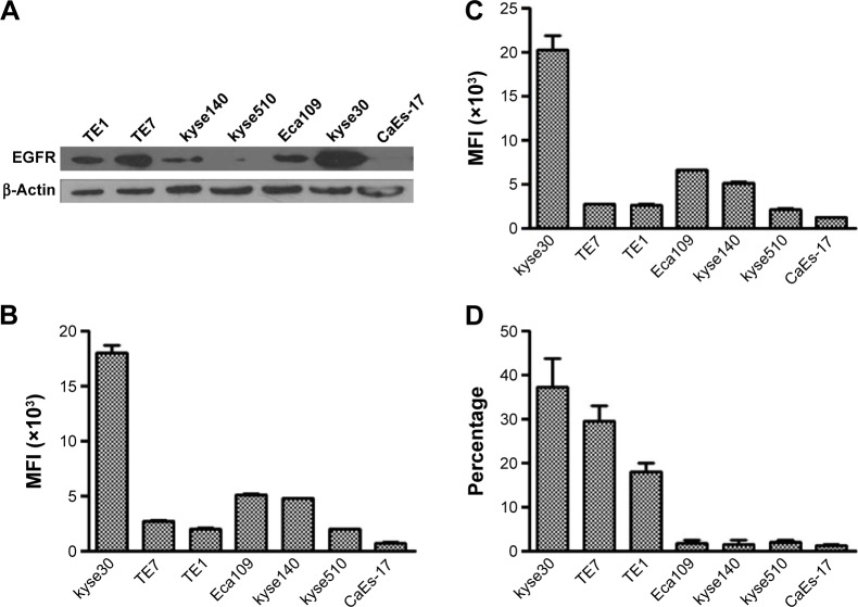
Purpose: The purpose of this study was to investigate the potential effect of activation of epidermal growth factor receptor (EGFR) signaling pathway on the expression of programmed death-ligand 1 (PD-L1) in esophageal squamous cell carcinoma (ESCC) cells with EGFR overexpression.
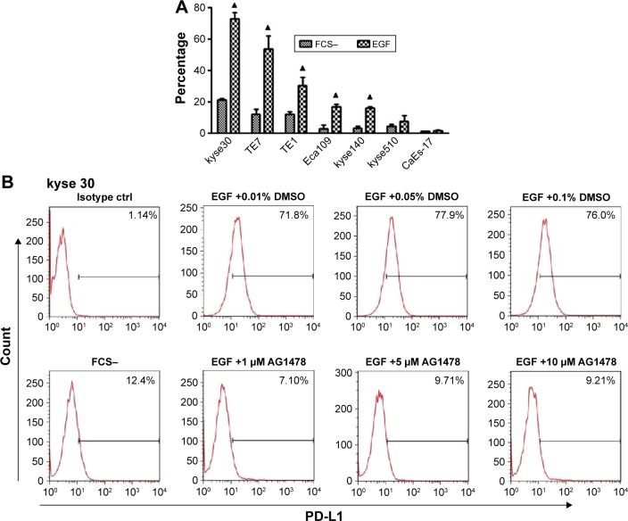
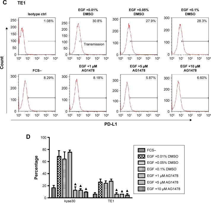
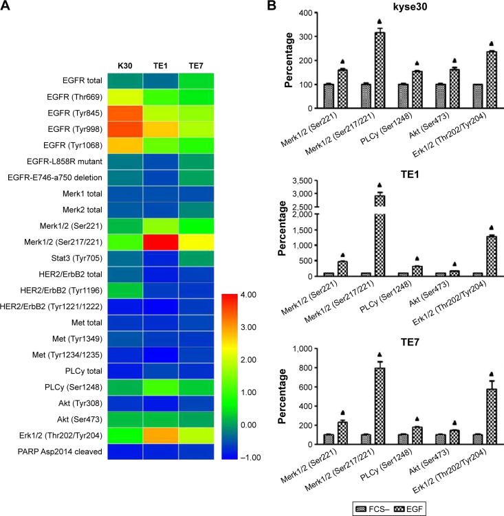
Methods: Flow cytometry and Western blot methods were used to assess PD-L1 expression on ESCC cells when EGFR signaling pathway was activated by epidermal growth factor (EGF) with or without EGFR-specific inhibitor AG-1478, and then EGFR signaling array was applied to analyze the potential signaling pathways involved.
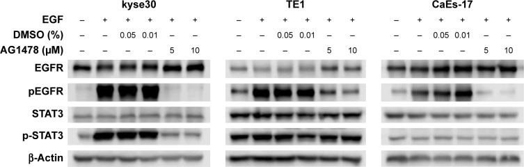
Results: This study found that PD-L1 expression increased significantly in an EGFR-dependent manner by the activation of EGFR signaling and decreased sharply when EGFR signaling was blocked. The upregulated expression of PD-L1 was not associated with EGFR-STAT3 signaling pathway, but may be affected by EGFR-PI3K-AKT, EGFR-Ras-Raf-Erk, and EGR-PLC-γ signaling pathways.
Conclusion: The expression of PD-L1 can be regulated by EGFR signaling activation in ESCC, which indicates an important role for EGFR-mediated immune escape and potential molecular pathways for EGFR-targeted therapy and immunotherapy.
Keywords: epidermal growth factor receptor; esophageal squamous cell carcinoma; immune checkpoint; programmed death-ligand 1.
Conflict of interest statement
Disclosure The authors report no conflicts of interest in this work.
Figures
Similar articles
-
Programmed death-ligand 1 is prognostic factor in esophageal squamous cell carcinoma and is associated with epidermal growth factor receptor.Cancer Sci. 2017 Apr;108(4):590-597. doi: 10.1111/cas.13197. Epub 2017 Apr 25. Cancer Sci. 2017. PMID: 28192623 Free PMC article.
-
Prognostic significance of epidermal growth factor receptor and programmed cell death-ligand 1 co-expression in esophageal squamous cell carcinoma.Aging (Albany NY). 2023 Feb 20;15(4):1107-1129. doi: 10.18632/aging.204535. Epub 2023 Feb 20. Aging (Albany NY). 2023. PMID: 36812484 Free PMC article.
-
Upregulation of PD-L1 by EGFR Activation Mediates the Immune Escape in EGFR-Driven NSCLC: Implication for Optional Immune Targeted Therapy for NSCLC Patients with EGFR Mutation.J Thorac Oncol. 2015 Jun;10(6):910-23. doi: 10.1097/JTO.0000000000000500. J Thorac Oncol. 2015. PMID: 25658629
-
Mutual connected IL-6, EGFR and LIN28/Let7-related mechanisms modulate PD-L1 and IGF upregulation in HNSCC using immunotherapy.Front Oncol. 2023 Apr 12;13:1140133. doi: 10.3389/fonc.2023.1140133. eCollection 2023. Front Oncol. 2023. PMID: 37124491 Free PMC article. Review.
-
Epidermal Growth Factor Receptor (EGFR) Pathway, Yes-Associated Protein (YAP) and the Regulation of Programmed Death-Ligand 1 (PD-L1) in Non-Small Cell Lung Cancer (NSCLC).Int J Mol Sci. 2019 Aug 5;20(15):3821. doi: 10.3390/ijms20153821. Int J Mol Sci. 2019. PMID: 31387256 Free PMC article. Review.
Cited by
-
Advances in immuno-oncology biomarkers for gastroesophageal cancer: Programmed death ligand 1, microsatellite instability, and beyond.World J Gastroenterol. 2018 Jul 7;24(25):2686-2697. doi: 10.3748/wjg.v24.i25.2686. World J Gastroenterol. 2018. PMID: 29991874 Free PMC article. Review.
-
Metformin Downregulates PD-L1 Expression in Esophageal Squamous Cell Catrcinoma by Inhibiting IL-6 Signaling Pathway.Front Oncol. 2021 Nov 22;11:762523. doi: 10.3389/fonc.2021.762523. eCollection 2021. Front Oncol. 2021. PMID: 34881181 Free PMC article.
-
EGFR may participate in immune evasion through regulation of B7‑H5 expression in non‑small cell lung carcinoma.Mol Med Rep. 2018 Oct;18(4):3769-3779. doi: 10.3892/mmr.2018.9361. Epub 2018 Aug 8. Mol Med Rep. 2018. PMID: 30106102 Free PMC article.
-
Characterization of a Trispecific PD-L1 Blocking Antibody That Exhibits EGFR-Conditional 4-1BB Agonist Activity.Antibodies (Basel). 2024 Apr 24;13(2):34. doi: 10.3390/antib13020034. Antibodies (Basel). 2024. PMID: 38804302 Free PMC article.
-
Immunotherapy in Gastroesophageal Cancers: Current Evidence and Ongoing Trials.Curr Treat Options Oncol. 2021 Sep 15;22(11):100. doi: 10.1007/s11864-021-00893-6. Curr Treat Options Oncol. 2021. PMID: 34524553 Review.
References
-
- Pennathur A, Gibson MK, Jobe BA, Luketich JD. Oesophageal carcinoma. Lancet. 2013;381(9864):400–412. - PubMed
-
- Ayyappan S, Prabhakar D, Sharma N. Epidermal growth factor receptor (EGFR)-targeted therapies in esophagogastric cancer. Anticancer Res. 2013;33(10):4139–4155. - PubMed
-
- Zhang W, Zhu H, Liu X, et al. Epidermal growth factor receptor is a prognosis predictor in patients with esophageal squamous cell carcinoma. Ann Thorac Surg. 2014;98(2):513–519. - PubMed
-
- Wang J, Yu JM, Jing SW, et al. Relationship between EGFR over-expression and clinicopathologic characteristics in squamous cell carcinoma of the esophagus: a meta-analysis. Asian Pac J Cancer Prev. 2014;15(14):5889–5893. - PubMed
LinkOut - more resources
Full Text Sources
Other Literature Sources
Research Materials
Miscellaneous
