Review
. 2017 Feb 13:11:24.
doi: 10.3389/fncel.2017.00024. eCollection 2017.

Glial Cells and Their Function in the Adult Brain: A Journey through the History of Their Ablation

Affiliations
Review

Glial Cells and Their Function in the Adult Brain: A Journey through the History of Their Ablation

Sarah Jäkel et al. Front Cell Neurosci. .

Abstract

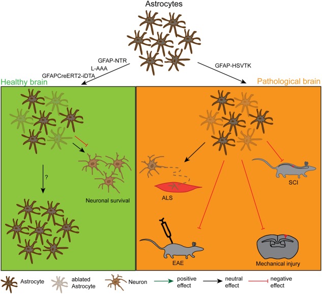
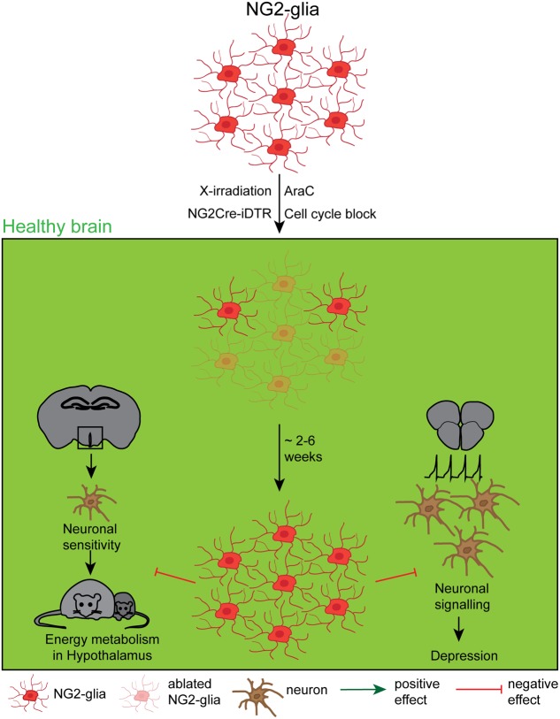
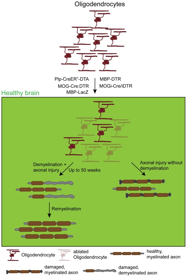
Glial cells, consisting of microglia, astrocytes, and oligodendrocyte lineage cells as their major components, constitute a large fraction of the mammalian brain. Originally considered as purely non-functional glue for neurons, decades of research have highlighted the importance as well as further functions of glial cells. Although many aspects of these cells are well characterized nowadays, the functions of the different glial populations in the brain under both physiological and pathological conditions remain, at least to a certain extent, unresolved. To tackle these important questions, a broad range of depletion approaches have been developed in which microglia, astrocytes, or oligodendrocyte lineage cells (i.e., NG2-glia and oligodendrocytes) are specifically ablated from the adult brain network with a subsequent analysis of the consequences. As the different glial populations are very heterogeneous, it is imperative to specifically ablate single cell populations instead of inducing cell death in all glial cells in general. Thanks to modern genetic manipulation methods, the approaches can now directly be targeted to the cell type of interest making the ablation more specific compared to general cell ablation approaches that have been used earlier on. In this review, we will give a detailed summary on different glial ablation studies, focusing on the adult mouse central nervous system and the functional readouts. We will also provide an outlook on how these approaches could be further exploited in the future.

Keywords: NG2-glia; astrocytes; brain function; cell ablation; microglia; oligodendrocytes.


Figures

FIGURE 1
FIGURE 1
Effects and dynamics of microglia ablation under healthy and pathological conditions. Under physiological conditions (left green panel) successful microglia ablation has been achieved by the use of the pharmacological inhibitors PLX3397 and clodronate liposomes (CLs) or the CX3CR1-iDTR mouse lines. After approximately 1 week, the microglia population was completely restored from a nestin+ progenitor pool. Depending on the study, the ablation had either no physiological effect or it influenced the motor-dependent synapse formation and led to a poor performance in learning tasks. Under various pathological conditions (right orange panel) the ablation was also induced with PLX3397, CL or different CD11b-HSVTK mouse lines, however, with very controversial outcomes. While the microglia ablation had positive effects on the pathology of experimental autoimmune encephalomyelitis (EAE) or the tauopathy of Alzheimer’s disease (AD), it did not influence the pathology of amyloid plaque deposition in AD, mechanical injury, amyotrophic lateral sclerosis (ALS), or temporal lobe epilepsy (TLE) and even negatively affected the outcome of a middle cerebral artery occlusion (MCAO).
FIGURE 2
FIGURE 2
Effects and dynamics of astrocyte ablation under healthy and pathological conditions. Under physiological conditions (left green panel) successful astrocyte ablation has been achieved by the use of the pharmacological drug L-α-aminoadipic acid (L-AAA) or the GFAPCreERT2-DTA and the GFAP-NTR mouse lines. For the astrocytes, the repopulation kinetics have not been analysed in detail yet, but these cells were also shown to repopulate the depleted area. As a functional outcome, the ablation did either not show an effect or had a negative effect on neuronal survival in the cerebellum and the spinal cord. Under pathological conditions (right orange panel) astrocyte ablation was solely achieved by the use of the GFAP-HSVTK mouse line. However, with this model only the pool of proliferating astrocytes can be depleted. The reduction of scar forming astrocytes in general had a negative outcome for the injury size and severity in spinal cord injury (SCI), mechanical brain injury and EAE. It did not affect the pathology in a model of ALS, but this could be due to the low amount of proliferating astrocytes in this model.
FIGURE 3
FIGURE 3
Effects and dynamics of NG2-glia ablation under healthy conditions. Although a relatively unknown cell type, several approaches to deplete NG2-glia under physiological conditions, including the mitotic blocker arabinofuranosyl cytidine (AraC), a genetic cell cycle block, X-irradiation and the NG2Cre/iDTR mouse line. The very efficient repopulation occurred either immediately or between 2 and 6 weeks after the ablation, depending on the study. Although the function of NG2-glia has long been a mystery, recent studies showed that the NG2-glia ablation negatively affects the leptin-dependent energy metabolism and leads to a depression like behavior.
FIGURE 4
FIGURE 4
Effects and dynamics of oligodendrocyte ablation under healthy conditions. Under physiological conditions successful oligodendrocyte ablation has been achieved by the use of several approaches: the PLP-CreERT-DTA, the MOG-Cre:DTR, the MBP-DTR, the MBP-LacZ, and the MOGi-Cre/iDTR mouse lines. Very commonly, although very diverse in the use of promoters and suicide genes, these approaches induced oligodendrocyte death that in most cases resulted in primary demyelination followed by secondary induced neuronal damage. These observations were in most cases accompanied by a behavioral phenotype resulting from demyelination. This phenotype could, however, take up to 50 weeks to appear, depending on the model. After a longer time, demyelination was followed by a spontaneous remyelination. Only one study observed axonal damage without global demyelination that could be due to the loss of trophic axonal support.

References

    1. Akiyama H., McGeer P. L. (1990). Brain microglia constitutively express beta-2 integrins. J. Neuroimmunol. 30 81–93. 10.1016/0165-5728(90)90055-R - DOI - PubMed
    1. Amat J. A., Ishiguro H., Nakamura K., Norton W. T. (1996). Phenotypic diversity and kinetics of proliferating microglia and astrocytes following cortical stab wounds. Glia 16 368–382. 10.1002/(SICI)1098-1136(199604)16:4<368::AID-GLIA9<3.0.CO;2-W - DOI - PubMed
    1. Asai H., Ikezu S., Tsunoda S., Medalla M., Luebke J., Haydar T., et al. (2015). Depletion of microglia and inhibition of exosome synthesis halt tau propagation. Nat. Neurosci. 18 1584–1593. 10.1038/nn.4132 - DOI - PMC - PubMed
    1. Azevedo F. A. C., Carvalho L. R. B., Grinberg L. T., Farfel J. M., Ferretti R. E. L., Leite R. E. P., et al. (2009). Equal numbers of neuronal and nonneuronal cells make the human brain an isometrically scaled-up primate brain. J. Comp. Neurol. 513 532–541. 10.1002/cne.21974 - DOI - PubMed
    1. Bardehle S., Krüger M., Buggenthin F., Schwausch J., Ninkovic J., Clevers H., et al. (2013). Live imaging of astrocyte responses to acute injury reveals selective juxtavascular proliferation. Nat. Neurosci. 16 580–586. 10.1038/nn.3371 - DOI - PubMed