Case Reports
. 2017:2017:3569512.
doi: 10.1155/2017/3569512. Epub 2017 Jan 24.

Lipoma Arborescens of the Knee: Report of Three Cases and Review of the Literature

Affiliations
Case Reports

Lipoma Arborescens of the Knee: Report of Three Cases and Review of the Literature

Ioannis Tsifountoudis et al. Case Rep Med. 2017.

Abstract

Lipoma arborescens is a chronic, slow-growing, intra-articular lesion of benign nature, which is characterized by villous proliferation of the synovium, with replacement of the subsynovial connective tissue by mature fat cells. It usually involves the suprapatellar pouch of the knee joint. It is not a neoplasm but is rather considered a nonspecific reactive response to chronic synovial irritation, due to either mechanical or inflammatory insults. We report three cases of lipoma arborescens affecting the knee, the first in a young male without previous history of arthritis or trauma, the second in a 58-year-old male associated with osteoarthritis, and the final in a 44-year-old male diagnosed with psoriatic arthritis, which cover the entire pathologic spectrum of this unusual entity. We highlight the clinical findings and imaging features, by emphasizing especially the role of MRI, in the differential diagnosis of other, more complex intra-articular masses.


Conflict of interest statement

The authors declare that there is no conflict of interests regarding the publication of this paper.

Figures

Figure 1
Figure 1
CT images on axial (a) and reconstructed coronal (b) plane in soft tissue window reveal a low-density intra-articular mass similar to fat with multiple villi projecting inwards (arrows), which are surrounded by joint fluid with higher density than fat but lower than water. Distention of the joint capsule is evident.
Figure 2
Figure 2
MR images on coronal T1-weighted (a), sagittal PD-weighted with fat saturation (FS) (b), and axial, sagittal, and coronal T1-weighted with FS after iv administration of gadolinium (c), (d), and (e), respectively, depict frond-like villi projecting inwards from the synovium with signal equal to fat on all imaging sequences (short arrows). After iv administration of gadolinium, enhancement of the overlying synovium is observed (long arrows), but no enhancement of the underlying fat is detected. Joint effusion is present distending the joint capsule, without any sign of menisci or ligaments' injury, or osseous erosions.
Figure 3
Figure 3
Open biopsy (intraoperative view). Incision is part of the final one (a). Biopsy specimen (b).
Figure 4
Figure 4
Open synovectomy (intraoperative view). Red-black region of synovium is the site of foregone biopsy (arrow).
Figure 5
Figure 5
Broad papillary projections lined by synovial membrane. The stroma consists of lobules of mature adipose tissue with focal chronic inflammatory infiltration [Hematoxylin and Eosin stain ×100 (a) and ×200 magnification (b)].
Figure 6
Figure 6
The patient in the first month postoperatively with his operated knee fully flexed.
Figure 7
Figure 7
MR images at the same sagittal level on T1-weighted (a), PD-weighted with FS (b), and T1-weighted with FS after iv administration of gadolinium (c) demonstrate medial compartment osteoarthritis with marginal osteophytes, chondral lesions, subchondral changes, and a complex tear of the posterior horn of medial meniscus. Mild villous frond-like fatty subsynovial hyperplasia is present in the suprapatellar bursa associated with synovial proliferation, which is enhanced after iv administration of gadolinium, and joint effusion (arrows). MR images at the same axial level on T1-weighted (d), PD-weighted with FS (e), and T1-weighted with FS after iv administration of gadolinium (f) confirm the above findings (arrows), which are strongly suggestive of LA.
Figure 8
Figure 8
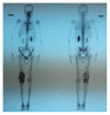
Tc-99m scintigraphy demonstrates persistent concentration of the technetium in the right knee joint, which is indicative of an inflammatory process.
Figure 9
Figure 9
US examination of the right knee on axial (a) and longitudinal (b) sonograms reveals hyperechoic frond-like projections of the synovium into the effusion (arrows), findings suggestive of LA. Colour Doppler US (c) does not depict any blood flow in the affected area.
Figure 10
Figure 10
MR images on sagittal T1-weighted (a) and PD-weighted with FS (b), on coronal T1-weighted (c) and PD-weighted with FS (d), and on axial STIR (e) and T2-weighted (f) demonstrate synovial proliferation and a large primarily fat-signal mass on all MR sequences, filling much of the suprapatellar bursa with a frond-like appearance, which is surrounded by excessive joint fluid. The innumerable individual villi are small in this case, with small quantities of interposed fluid, creating a feathery appearance (arrows). These findings are indicative of a large feathery-appearing LA filling the suprapatellar recess.

References

    1. Kloen P., Keel S. B., Chandler H. P., Geiger R. H., Zarins B., Rosenberg A. E. Lipoma arborescens of the knee. Journal of Bone and Joint Surgery B. 1998;80(2):298–301. doi: 10.1302/0301-620x.80b2.8244. - DOI - PubMed
    1. Sanamandra S. K., Ong K. O. Lipoma arborescens. Singapore Medical Journal. 2014;55(1):5–11. doi: 10.11622/smedj.2014003. - DOI - PMC - PubMed
    1. Kamran F., Kavin K., Vijay S., Shivanand G. Bilateral lipoma arborescens with osteoarthritis knee: case report and literature review. Journal of Clinical Orthopaedics and Trauma. 2015;6(2):131–136. doi: 10.1016/j.jcot.2014.12.006. - DOI - PMC - PubMed
    1. Arzimanoglu A. Bilateral arborescent lipoma of the knee. The Journal of Bone and Joint Surgery. 1957;39(4):976–979. - PubMed
    1. Kamaci S., Doral M. N., Ergen F. B., Yucekul A., Cil A. Lipoma arborescens of the knee. Knee Surgery, Sports Traumatology, Arthroscopy. 2015;23(8):2196–2201. doi: 10.1007/s00167-014-2996-3. - DOI - PubMed

Publication types

LinkOut - more resources