

Repeated Administration of Mercury Intensifies Brain Damage in Multiple Sclerosis through Mitochondrial Dysfunction
- PMID: 28243280
- PMCID: PMC5316262
Repeated Administration of Mercury Intensifies Brain Damage in Multiple Sclerosis through Mitochondrial Dysfunction
Abstract
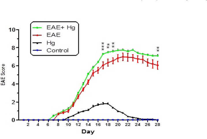
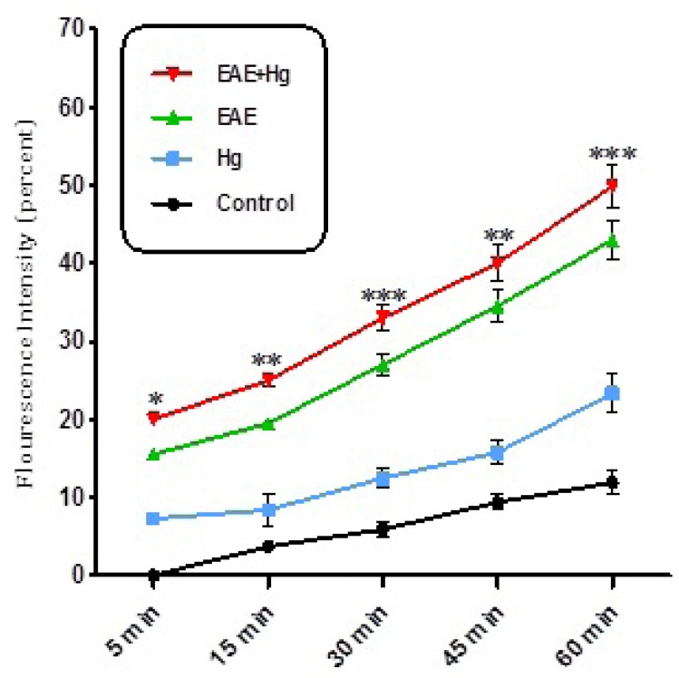
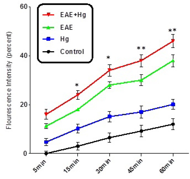
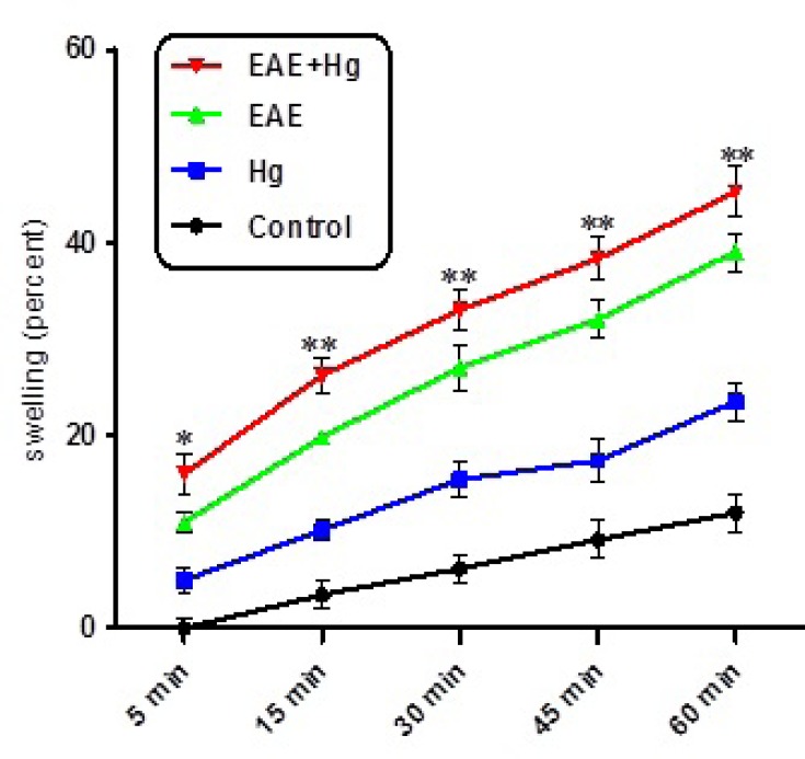
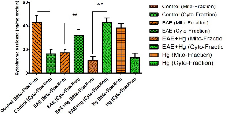
In this study we investigated the additive effect of mercury on the brain mitochondrial dysfunction in experimental autoimmune encephalomyelitis (EAE) model. Experimental animals (female C57BL/6 mice) are divided into four groups (n = 8); control, Hg, EAE, EAE with Hg. EAE model of MS induced by injecting myelin oligodendrocyte glycoprotein (MOG). Neurobehavioral alterations are recorded and then mice were sacrificed at day 28 and brain mitochondria were isolated and mitochondrial toxicity parameters including mitochondrial swelling, reactive oxygen species (ROS) formation, collapse of mitochondrial membrane potential (MMP) and cytochrome c release were measured. Our results showed that repeated treatment of mercury following induction of EAE in mice significantly increased the neurobehavioral scores, as well as mitochondrial toxicity through ROS formation, mitochondrial swelling, collapse of MMP and cytochrome c release. Our findings proved that repeated exposure with mercury accelerates progression of MS through mitochondrial damage related to oxidative stress and finally apoptosis.
Keywords: Apoptosis; Brain mitochondria; EAE model; Mercury; Oxidative stress.
Figures
References
-
- Gochfeld M. Cases of mercury exposure, bioavailability, and absorption. Ecotoxicol. Environ. Saf. 2003;56:174–9. - PubMed
-
- Hylander LD, Meili M. 500 years of mercury production: Global annual inventory by region until 2000 and associated emissions. Sci. Total Environ. 2003;304:13–27. - PubMed
-
- Moreau T, Coles A, Wing M, Isaacs J, Hale G, Waldmann H, Compston A. Transient increase in symptoms associated with cytokine release in patients with multiple sclerosis. Brain. 1996;119:225–38. - PubMed
-
- Beal M. Aging, energy, and oxidative stress in neurodegenerative diseases. Restor. Neurol. Neurosci. 1996;3:180–1. - PubMed
LinkOut - more resources
Full Text Sources