

A Modified Method for Creating Elastase-Induced Aneurysms by Ligation of Common Carotid Arteries in Rabbits and Its Effect on Surrounding Arteries
- PMID: 28243348
- PMCID: PMC5317289
A Modified Method for Creating Elastase-Induced Aneurysms by Ligation of Common Carotid Arteries in Rabbits and Its Effect on Surrounding Arteries
Abstract
Background and purpose: Rabbit models of intracranial aneurysms are frequently used in pre-clinical settings. This study aimed to demonstrate an alternative, extravascular method for creating elastase-induced aneurysms, and how ligation of the right common carotid arteries (RCCA) can impact flow redistribution into left CCA (LCCA).
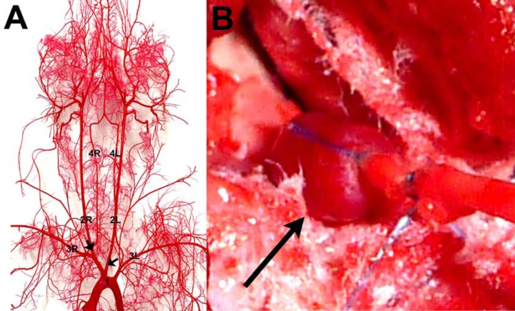
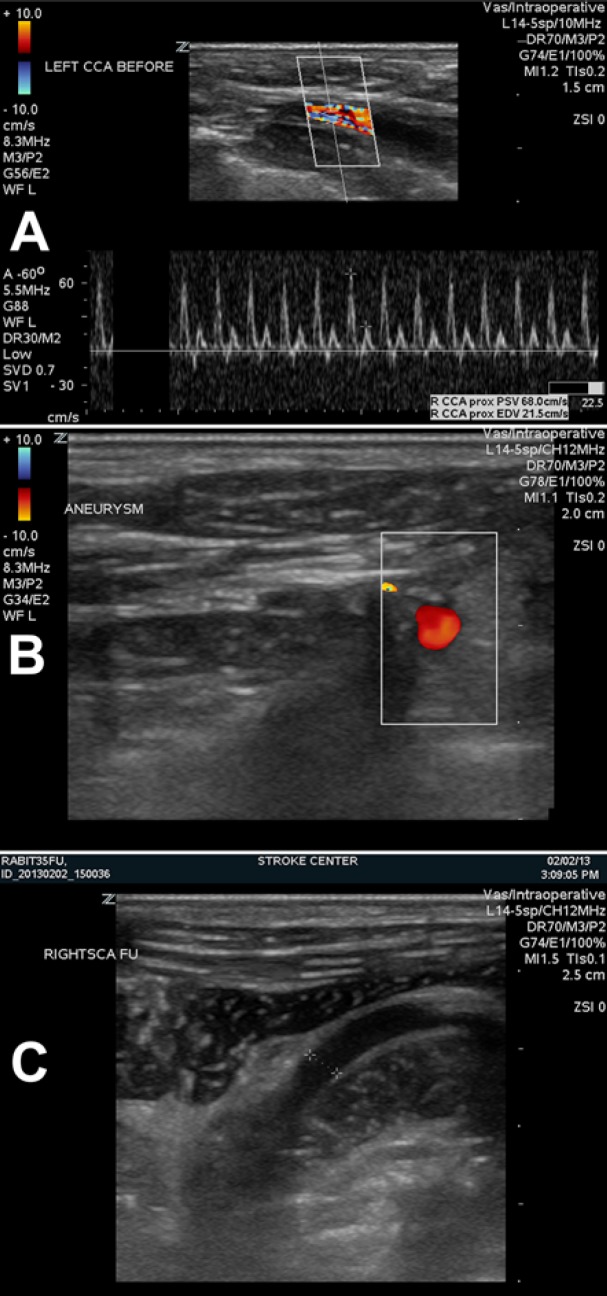
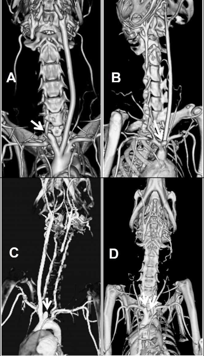
Methods: Elastase-induced aneurysms in 18 New Zealand rabbits (4.14 ± 0.314 kg) were created by applying 3-5 U of concentrated elastase solution to the exterior of the right and left CCA roots (RCCA and LCCA). After the induction of the aneurysm, the aneurysm was either kept intact to the rest of the corresponding CCA, severed from the rest of the CCA to allow for a free standing aneurysm, or was anchored to nearby tissue to influence the angle and orientation of the aneurysm with respect to the parent vessel. Ultrasound studies were performed before and after creation of aneurysms to collect blood flow measurements inside the aneurysm pouch and surrounding arteries. Prior to sacrificing the animals, computed tomography angiography studies were performed. Harvested aneurysmal tissues were used for histological analysis.
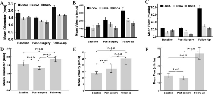
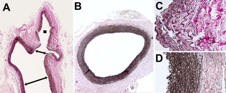
Results: Elastase-induced aneurysms were successfully created by the extravascular approach. Histological studies showed that the biological response was similar to human cerebral aneurysms and previously published elastase-induced rabbit aneurysm models. Ultrasound measurements indicated that after the RCCA was ligated, blood flow significantly increased in the LCCA at one-month follow-up.
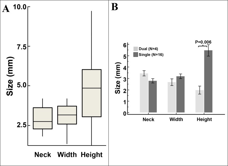
Conclusion: An alternate method for creating elastase-induced aneurysms has been demonstrated. The novel aspects of our method allow for ligation of one or both common carotid arteries to create a single or bilateral aneurysm with an ability to control the orientation of the induced aneurysm.
Keywords: aneurysm model; angiography; elastase; flow diverter; rabbit model; ultrasound.
Figures
Similar articles
-
An Effective and Simple Way to Establish Elastase-Induced Middle Carotid Artery Fusiform Aneurysms in Rabbits.Biomed Res Int. 2020 Aug 26;2020:6707012. doi: 10.1155/2020/6707012. eCollection 2020. Biomed Res Int. 2020. PMID: 32908904 Free PMC article.
-
A modified technique for using elastase to create saccular aneurysms in animals that histologically and hemodynamically resemble aneurysms in human.Acta Neurochir (Wien). 2004 Jul;146(7):705-11. doi: 10.1007/s00701-004-0276-6. Epub 2004 May 17. Acta Neurochir (Wien). 2004. PMID: 15197614
-
Serial angiography in an elastase-induced aneurysm model in rabbits: evidence for progressive aneurysm enlargement after creation.AJNR Am J Neuroradiol. 2001 Apr;22(4):698-703. AJNR Am J Neuroradiol. 2001. PMID: 11290481 Free PMC article.
-
Creation of Two Saccular Elastase-Digested Aneurysms with Different Hemodynamics in One Rabbit.J Vis Exp. 2021 Apr 15;(170). doi: 10.3791/62518. J Vis Exp. 2021. PMID: 33938900
-
Preclinical Intracranial Aneurysm Models: A Systematic Review.Brain Sci. 2020 Feb 27;10(3):134. doi: 10.3390/brainsci10030134. Brain Sci. 2020. PMID: 32120907 Free PMC article. Review.
Cited by
-
An Effective and Simple Way to Establish Elastase-Induced Middle Carotid Artery Fusiform Aneurysms in Rabbits.Biomed Res Int. 2020 Aug 26;2020:6707012. doi: 10.1155/2020/6707012. eCollection 2020. Biomed Res Int. 2020. PMID: 32908904 Free PMC article.
-
Preclinical extracranial aneurysm models for the study and treatment of brain aneurysms: A systematic review.J Cereb Blood Flow Metab. 2020 May;40(5):922-938. doi: 10.1177/0271678X20908363. Epub 2020 Mar 3. J Cereb Blood Flow Metab. 2020. PMID: 32126875 Free PMC article.
-
Imaging Modalities for Intracranial Aneurysm: More Than Meets the Eye.Front Cardiovasc Med. 2022 Feb 15;9:793072. doi: 10.3389/fcvm.2022.793072. eCollection 2022. Front Cardiovasc Med. 2022. PMID: 35242823 Free PMC article. Review.
References
-
- van Donkelaar CE, et al. Predictive factors for rebleeding after aneurysmal subarachnoid hemorrhage: rebleeding aneurysmal subarachnoid hemorrhage study. Stroke: a journal of cerebral circulation. 2015;46(8):2100–2106. - PubMed
-
- Finlay HM, et al. Collagen organization in the branching region of human brain arteries. Stroke: a journal of cerebral circulation. 1998;29(8):1595–1601. - PubMed
LinkOut - more resources
Full Text Sources