

Pityriasis lichenoides et varioliformis acuta in skin of color: new observations by dermoscopy
- PMID: 28243491
- PMCID: PMC5315037
- DOI: 10.5826/dpc.0701a05
Pityriasis lichenoides et varioliformis acuta in skin of color: new observations by dermoscopy
Abstract
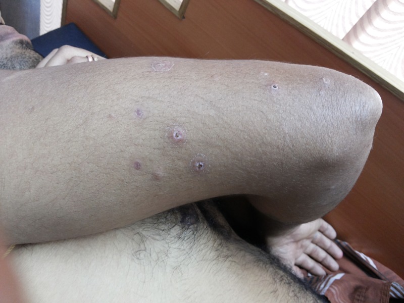
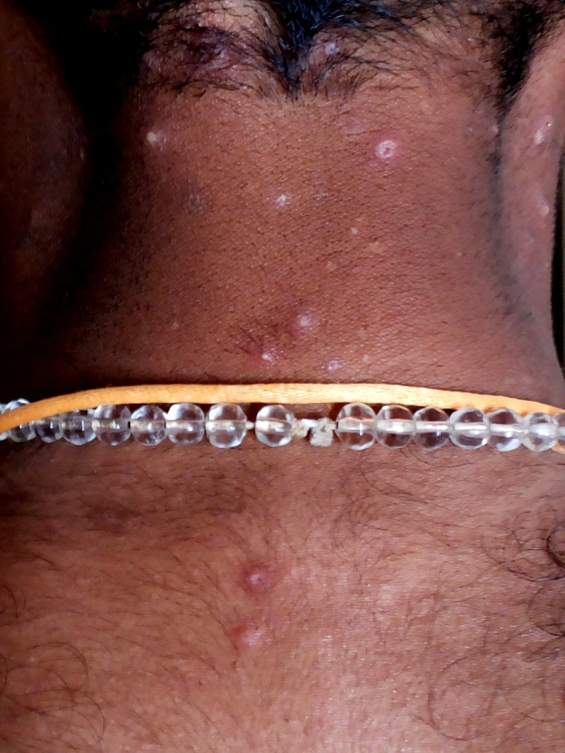
Background: Pityriasis lichenoides is an uncommon skin disease that presents in three different forms: pityriasis lichenoides et varioliformis acuta (PLEVA), pityriasis lichenoides chronica (PLC) and febrile ulceronecrotic-Mucha-Habermann disease. These represent a spectrum of a disease. PLEVA presents as skin eruption of multiple, small, red papules that develop into polymorphic lesions with periods of varying remissions, as well as possible sequels of hyper/hypopigmentation and varicella-like scars. Diagnosis of this condition is mainly clinical, and sometimes clinical differentiation from other conditions may be a difficult task that often requires histological analysis. In this study, PLEVA lesions were examined by dermoscopy, and the significance of specific dermoscopic findings was investigated in order to facilitate their differentiation from other inflammatory conditions.
Objectives: To evaluate dermoscopic patterns in PLEVA and to correlate these patterns with histopathology.
Materials and methods: The study was conducted at S. Nijalingappa Medical College, Bagalkot. It was an observational case series study and patients were selected randomly. Ethical clearance and informed consent were obtained. PLEVA lesions in early and late phases were evaluated. A manual DermLite 3 (3Gen, San Juan Capistrano, CA) dermoscope attached to a Sony (Cyber Shot DSC-W800, Sony Electronics Inc., San Diego, California, USA, digital, 14 mega pixels) camera was employed. Histopathology was done to confirm the diagnosis. Data was collected and analyzed. Results were statistically described in terms of frequencies and types of dermoscopic patterns.
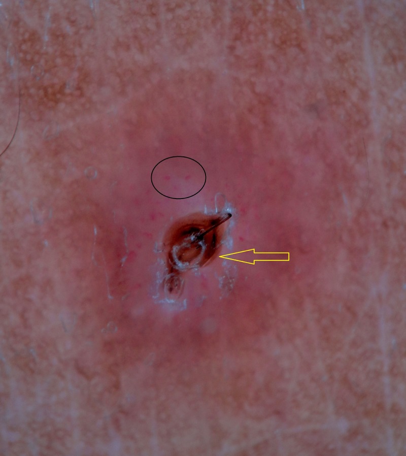
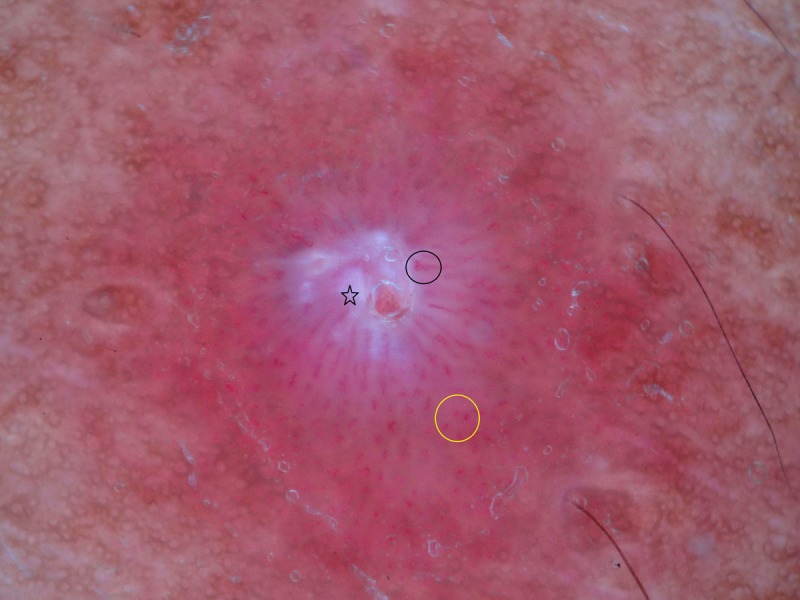
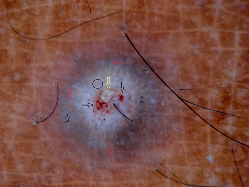
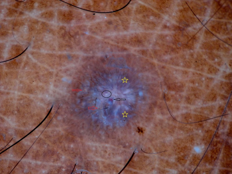
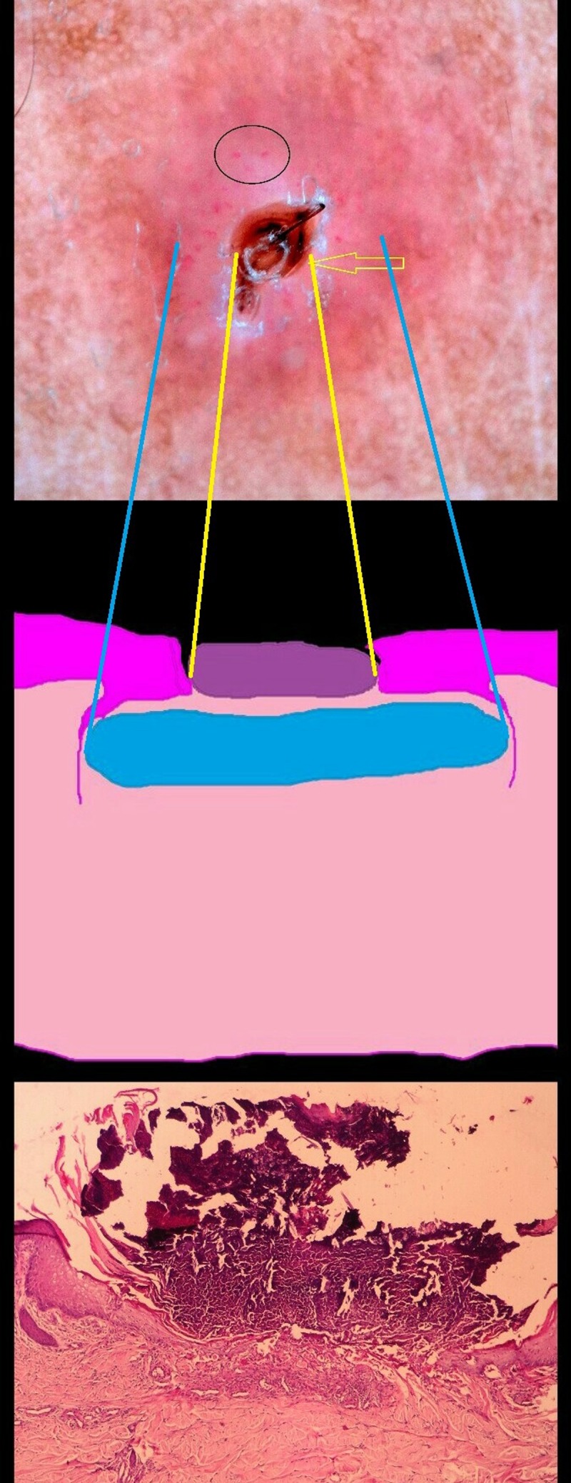
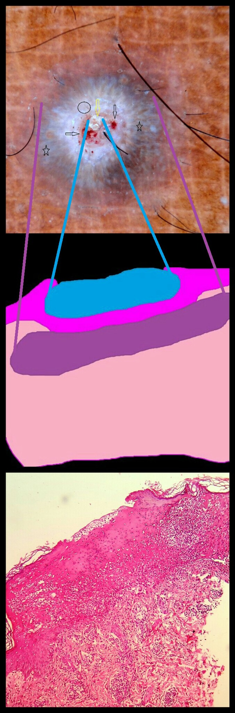
Results: There was a total of 14 patients; 8 males and 6 females. Mean age of patients was 19 years. Mean duration of disease was 7 months. Dermoscopy in early-phase lesions revealed amorphous brownish areas around the hair follicles, dotted vessels, and scaling. Dermoscopy in late-phase lesions showed whitish-structureless areas and central white crust within whitish-structureless rim with scale, focal bluish-grayish areas or centrifugal strands irregularly distributed along the periphery and yellow structures. Red dots and hemorrhage were seen at the center and glomerular vessels at the periphery.
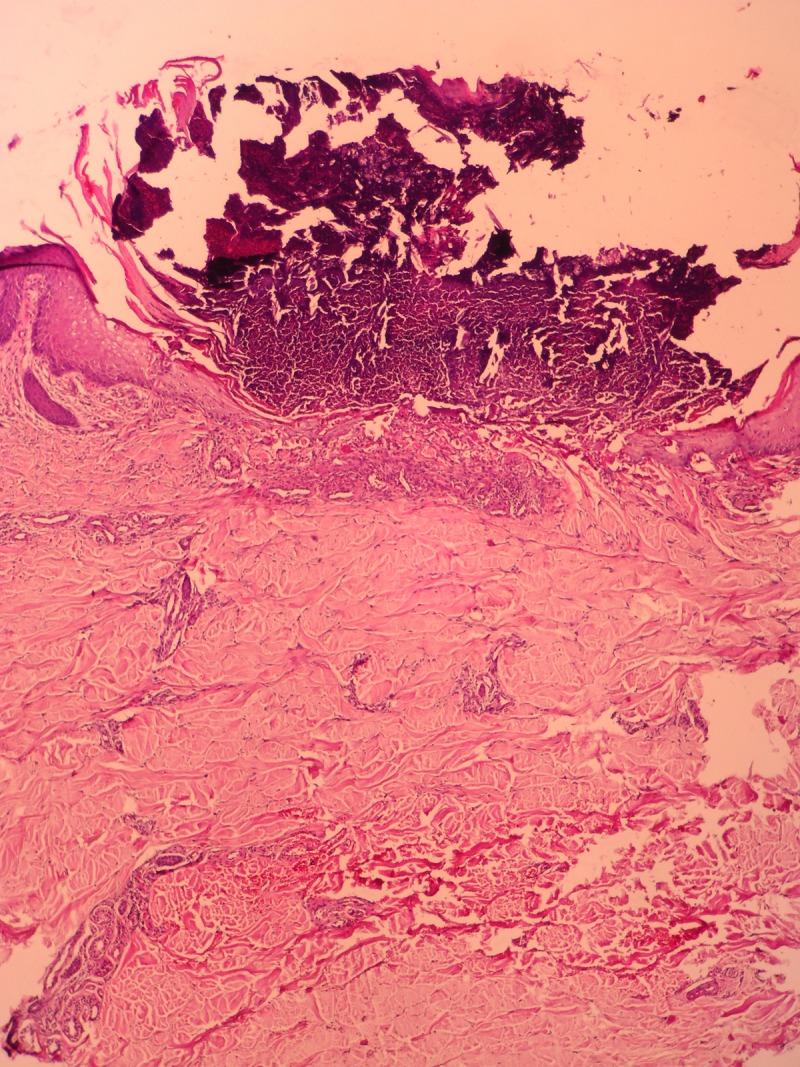
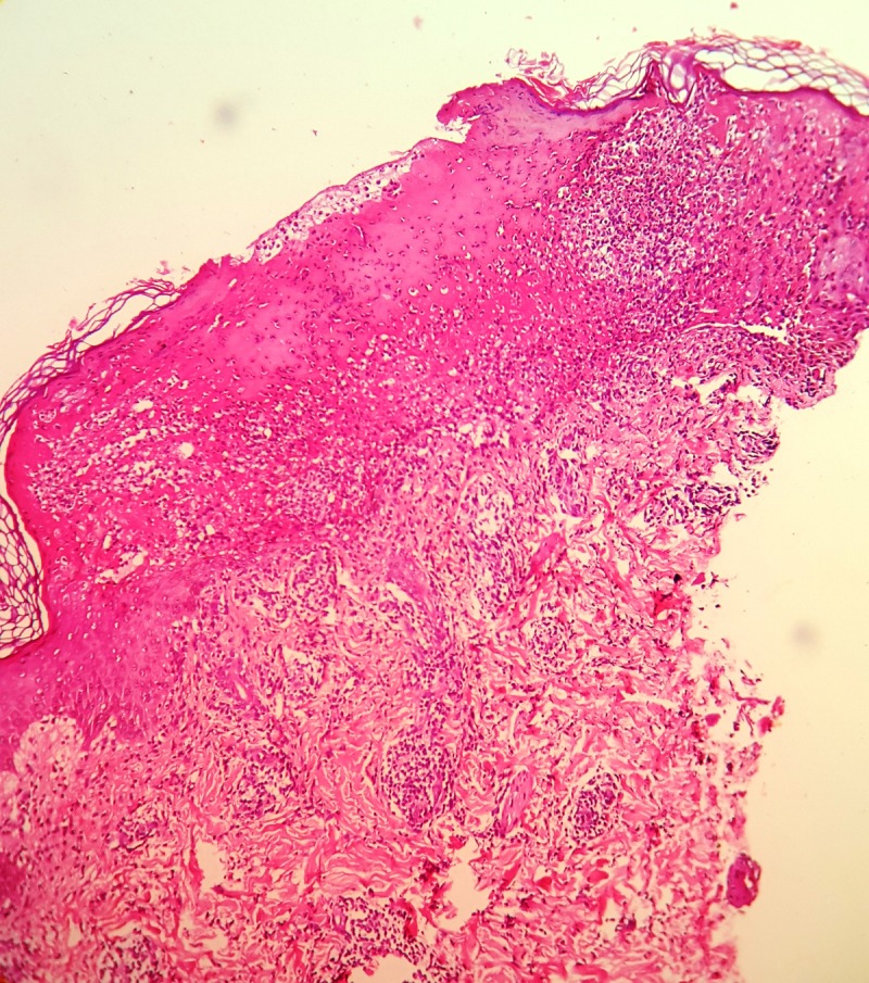
Conclusion: PLEVA demonstrates specific dermoscopic patterns that correlate well with histologic changes. New dermoscopic findings are described. Thus, dermoscopy is a good diagnostic tool in the clinical diagnosis of PLEVA.
Keywords: dermoscopy; diagnosis; pattern; pityriasis lichenoides et varioliformis acuta.
Conflict of interest statement
Competing interests: The authors have no conflicts of interest to disclose.
Figures
Similar articles
-
Pityriasis lichenoides: pathophysiology, classification, and treatment.Am J Clin Dermatol. 2007;8(1):29-36. doi: 10.2165/00128071-200708010-00004. Am J Clin Dermatol. 2007. PMID: 17298104 Review.
-
Hypertrophic lichen planus versus prurigo nodularis: a dermoscopic perspective.Dermatol Pract Concept. 2016 Apr 30;6(2):9-15. doi: 10.5826/dpc.0602a03. eCollection 2016 Apr. Dermatol Pract Concept. 2016. PMID: 27222766 Free PMC article.
-
Clinical, Dermatoscopic, and Histological Findings in a Diagnosis of Pityriasis Lichenoides.Cureus. 2020 Jun 20;12(6):e8725. doi: 10.7759/cureus.8725. Cureus. 2020. PMID: 32699720 Free PMC article.
-
Oral erythromycin in pityriasis lichenoides chronica and pityriasis lichenoides et varioliformis acuta.Dermatol Ther. 2020 May;33(3):e13311. doi: 10.1111/dth.13311. Epub 2020 Mar 30. Dermatol Ther. 2020. PMID: 32174014
-
Pityriasis lichenoides and its subtypes.J Am Acad Dermatol. 2006 Oct;55(4):557-72; quiz 573-6. doi: 10.1016/j.jaad.2005.07.058. J Am Acad Dermatol. 2006. PMID: 17010734 Review.
Cited by
-
Dermatoscopy in Skin of Color: How Different are We?Indian Dermatol Online J. 2021 Jan 16;12(1):12-13. doi: 10.4103/idoj.IDOJ_625_20. eCollection 2021 Jan-Feb. Indian Dermatol Online J. 2021. PMID: 33768018 Free PMC article. No abstract available.
-
Dermoscopy of pityriasis lichenoides et varioliformis acuta (PLEVA).An Bras Dermatol. 2024 Jan-Feb;99(1):120-123. doi: 10.1016/j.abd.2022.04.017. Epub 2023 Sep 1. An Bras Dermatol. 2024. PMID: 37661463 Free PMC article. No abstract available.
-
Dermatoscopy of Inflammatory Diseases in Skin of Color.Indian Dermatol Online J. 2021 Jan 16;12(1):45-57. doi: 10.4103/idoj.IDOJ_613_20. eCollection 2021 Jan-Feb. Indian Dermatol Online J. 2021. PMID: 33768022 Free PMC article. Review.
-
Dermoscopy of Inflammatory Dermatoses (Inflammoscopy) in Skin of Color - A Systematic Review by the International Dermoscopy Society "Imaging in Skin of Color" Task Force.Dermatol Pract Concept. 2023 Oct 1;13(4 S1):e2023297S. doi: 10.5826/dpc.1304S1a297S. Dermatol Pract Concept. 2023. PMID: 37874994 Free PMC article. Review.
-
Diagnostic Dermoscopy in Pityriasis Lichenoides Chronica and Pityriasis Lichenoides et Varioliformis Acuta: A Case Series.Dermatol Pract Concept. 2025 Apr 1;15(2):5078. doi: 10.5826/dpc.1502a5078. Dermatol Pract Concept. 2025. PMID: 40401893 Free PMC article. No abstract available.
References
-
- Perrin BS, Yan AC, Treat JR. Febrile ulceronecrotic Mucha-Habermann disease in a 34-month-old boy: a case report and review of the literature. Paediatric Dermatol. 2012;29(1):53–58. - PubMed
-
- Khachemoune A, Blyumin ML. Pityriasis lichenoides: pathophysiology, classification, and treatment. Am J Clin Dermatol. 2007;8(1):29–36. - PubMed
-
- Errichetti E, Stinco G. The practical usefulness of dermoscopy in general dermatology. G Ital Dermatol Venereol. 2015;150(5):533–546. - PubMed
-
- Bowling J. Introduction to dermoscopy. In: Bowling J, editor. Diagnostic Dermoscopy: The Illustrated Guide. 1st ed. West Sussex: Wiley-Blackwell; 2012. pp. 2–14.
-
- Kreusch J. How to perform dermoscopy of non-pigmented skin lesions. In: Zalaudek I, Argenziano G, Giacomel J, editors. Dermatoscopy of Non-Pigmented Skin Tumors: Pink – Think - Blink. New York: CRC Press; 2016. pp. 17–18.
LinkOut - more resources
Full Text Sources
Other Literature Sources