

Dermoscopic and confocal features of an axillary "special site" nevus
- PMID: 28243497
- PMCID: PMC5315043
- DOI: 10.5826/dpc.0701a11
Dermoscopic and confocal features of an axillary "special site" nevus
Abstract
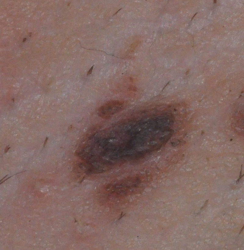
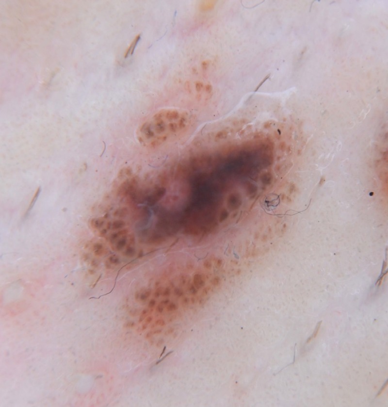
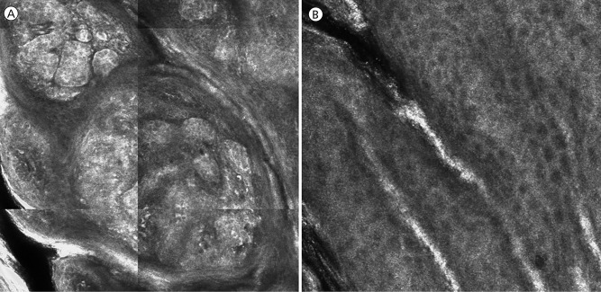
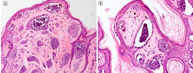
"Nevi of special sites" is a term that denotes melanocytic nevi presenting in specific anatomic locations including the scalp, genital area, flexural sites, and acral sites [1]. Nevi from these anatomic sites display at times histopathologic features that may lead the reading pathologist to recommend re-excision of these benign nevi. Reflectance confocal microscopy (RCM) is a noninvasive imaging tool that allows for visualization of epidermal, dermal-epidermal junctional (DEJ), and superficial dermal tissue structures at cellular level resolution. RCM features of special site nevi have not been previously described in the literature. Defining the RCM characteristics of special site nevi may increase diagnostic accuracy and assist in ruling out melanoma. Here, we report a case of a pigmented lesion appearing in the axilla of a patient with a recently diagnosed melanoma. Dermoscopic and histopathologic results were consistent with the diagnosis of nevus in flexural anatomic sites. In this case, RCM showed a regular honeycomb pattern of epidermal keratinocytes and enlarged, non-homogenous, discohesive nests at the DEJ, a pattern that corresponded well with the histopathologic findings. Larger studies are needed to establish RCM features of special site nevi in order to reliably rule out melanoma and lower the rate of unnecessary excisions of these benign nevi.
Conflict of interest statement
Competing interests: The authors have no conflicts of interest to disclose.
Figures
Similar articles
-
Role of In Vivo Reflectance Confocal Microscopy in the Analysis of Melanocytic Lesions.Acta Dermatovenerol Croat. 2018 Apr;26(1):64-67. Acta Dermatovenerol Croat. 2018. PMID: 29782304 Review.
-
Nested Melanoma, a New Morphological Variant of Superficial Spreading Melanoma with Characteristic Dermoscopic Features.Acta Dermatovenerol Croat. 2017 Apr;25(1):80-81. Acta Dermatovenerol Croat. 2017. PMID: 28511756
-
Nevomelanocytic atypia detection by in vivo reflectance confocal microscopy.Medicina (Kaunas). 2014;50(4):209-15. doi: 10.1016/j.medici.2014.09.008. Epub 2014 Oct 1. Medicina (Kaunas). 2014. PMID: 25458957
-
Reflectance confocal microscopy analysis of equivocal melanocytic lesions with severe regression.Skin Res Technol. 2018 Feb;24(1):9-15. doi: 10.1111/srt.12382. Epub 2017 May 21. Skin Res Technol. 2018. PMID: 28543606
-
Melanocytic nevi with special features: clinical-dermoscopic and reflectance confocal microscopic-findings.J Eur Acad Dermatol Venereol. 2014 Jul;28(7):833-45. doi: 10.1111/jdv.12291. Epub 2013 Oct 31. J Eur Acad Dermatol Venereol. 2014. PMID: 24171788 Review.
References
-
- Mason AR, Mohr MR, Koch LH, Hood AF. Nevi of special sites. Clin Lab Med. 2011;31(2):229–242. - PubMed
-
- Buonaccorsi JN, Lynott J, Plaza JA. Atypical melanocytic lesions of the thigh with spitzoid and dysplastic features: A clinicopathologic study of 29 cases. Ann Diagn Pathol. 2013;17(3):265–269. - PubMed
-
- Hofmann-Wellenhof R. Special criteria for special locations: Scalp, mucosal, and milk line. Dermatol Clin. 2013;31(4):625–636. - PubMed
-
- Massi G, LeBoit PE. Histological Diagnosis of Nevi and Melanoma. Darmstadt: Steinkopff Verlag; 2004. Polypoid nevus of pregnancy and milk line nevi; pp. 329–336.
-
- Merkel EA, Martini MC, Amin SM, Lee CY, Gerami P. Evaluation of dermoscopic features for distinguishing melanoma from special site nevi of the breast. J Am Acad Dermatol. 2016;75(2):1–7. - PubMed
LinkOut - more resources
Full Text Sources
Other Literature Sources