Development of novel murine mammary imaging windows to examine wound healing effects on leukocyte trafficking in mammary tumors with intravital imaging
- PMID: 28243517
- PMCID: PMC5226013
- DOI: 10.1080/21659087.2015.1125562
Development of novel murine mammary imaging windows to examine wound healing effects on leukocyte trafficking in mammary tumors with intravital imaging
Abstract
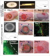
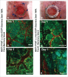
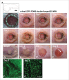
We developed mammary imaging windows (MIWs) to evaluate leukocyte infiltration and cancer cell dissemination in mouse mammary tumors imaged by confocal microscopy. Previous techniques relied on surgical resection of a skin flap to image the tumor microenvironment restricting imaging time to a few hours. Utilization of mammary imaging windows offers extension of intravital imaging of the tumor microenvironment. We have characterized strengths and identified some previously undescribed potential weaknesses of MIW techniques. Through iterative enhancements of a transdermal portal we defined conditions for improved quality and extended confocal imaging time for imaging key cell-cell interactions in the tumor microenvironment.
Keywords: breast cancer; cell migration; intravital imaging; mammary imaging windows.
Figures


Similar articles
-
Longitudinal Intravital Microscopy Using a Mammary Imaging Window with Replaceable Lid.J Vis Exp. 2022 Jan 20;(179). doi: 10.3791/63326. J Vis Exp. 2022. PMID: 35129183
-
Dendra2 photoswitching through the Mammary Imaging Window.J Vis Exp. 2009 Jun 5;(28):1278. doi: 10.3791/1278. J Vis Exp. 2009. PMID: 19578330 Free PMC article.
-
Tracking Cell Recruitment and Behavior within the Tumor Microenvironment Using Advanced Intravital Imaging Approaches.Cells. 2018 Jul 3;7(7):69. doi: 10.3390/cells7070069. Cells. 2018. PMID: 29970845 Free PMC article.
-
Procedures and applications of long-term intravital microscopy.Methods. 2017 Sep 1;128:52-64. doi: 10.1016/j.ymeth.2017.06.029. Epub 2017 Jun 30. Methods. 2017. PMID: 28669866 Review.
-
In Vivo Imaging Sheds Light on Immune Cell Migration and Function in Cancer.Front Immunol. 2017 Mar 22;8:309. doi: 10.3389/fimmu.2017.00309. eCollection 2017. Front Immunol. 2017. PMID: 28382036 Free PMC article. Review.
Cited by
-
Longitudinal Intravital Imaging Through Clear Silicone Windows.J Vis Exp. 2022 Jan 5;(179):10.3791/62757. doi: 10.3791/62757. J Vis Exp. 2022. PMID: 35068483 Free PMC article.
-
Tools and Concepts for Interrogating and Defining Cellular Identity.Cell Stem Cell. 2020 May 7;26(5):632-656. doi: 10.1016/j.stem.2020.03.015. Cell Stem Cell. 2020. PMID: 32386555 Free PMC article. Review.
-
Intravital microscopy of dynamic single-cell behavior in mouse mammary tissue.Nat Protoc. 2021 Apr;16(4):1907-1935. doi: 10.1038/s41596-020-00473-2. Epub 2021 Feb 24. Nat Protoc. 2021. PMID: 33627843
-
Longitudinal intravital microscopy of the mouse kidney: inflammatory responses to abdominal imaging windows.Am J Physiol Renal Physiol. 2024 Nov 1;327(5):F845-F868. doi: 10.1152/ajprenal.00071.2024. Epub 2024 Sep 26. Am J Physiol Renal Physiol. 2024. PMID: 39323386
-
Visualizing vasculature and its response to therapy in the tumor microenvironment.Theranostics. 2023 Sep 25;13(15):5223-5246. doi: 10.7150/thno.84947. eCollection 2023. Theranostics. 2023. PMID: 37908739 Free PMC article. Review.
References
-
- Muller A, Homey B, Soto H, Ge N, Catron D, Buchanan ME, McClanahan T, Murphy E, Yuan W, Wagner SN, et al. . Involvement of chemokine receptors in breast cancer metastasis. Nature 2001; 410:50-6; PMID:11242036; http://dx.doi.org/10.1038/35065016 - DOI - PubMed
-
- Zlotnik A, Burkhardt AM, Homey B. Homeostatic chemokine receptors and organ-specific metastasis. Nat Rev Immunol 2011; 11:597-606; PMID:21866172; http://dx.doi.org/10.1038/nri3049 - DOI - PubMed
-
- Kato M, Kitayama J, Kazama S, Nagawa H. Expression pattern of CXC chemokine receptor-4 is correlated with lymph node metastasis in human invasive ductal carcinoma. Breast Cancer Res 2003; 5:R144-50; PMID:12927045; http://dx.doi.org/10.1186/bcr627 - DOI - PMC - PubMed
-
- Su YC, Wu MT, Huang CJ, Hou MF, Yang SF, Chai CY. Expression of CXCR4 is associated with axillary lymph node status in patients with early breast cancer. Breast 2006; 15:533-9; PMID:16239110; http://dx.doi.org/10.1016/j.breast.2005.08.034 - DOI - PubMed
-
- Kang H, Watkins G, Douglas-Jones A, Mansel RE, Jiang WG. The elevated level of CXCR4 is correlated with nodal metastasis of human breast cancer. Breast 2005; 14:360-7; PMID:16216737; http://dx.doi.org/10.1016/j.breast.2004.12.007 - DOI - PubMed
Grants and funding
LinkOut - more resources
Full Text Sources
Other Literature Sources
