

Inhibition of calcineurin by FK506 stimulates germinal vesicle breakdown of mouse oocytes in hypoxanthine-supplemented medium
- PMID: 28243539
- PMCID: PMC5326542
- DOI: 10.7717/peerj.3032
Inhibition of calcineurin by FK506 stimulates germinal vesicle breakdown of mouse oocytes in hypoxanthine-supplemented medium
Abstract
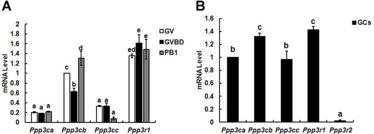
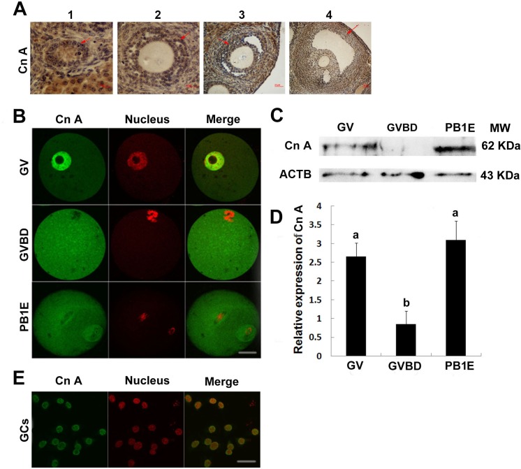
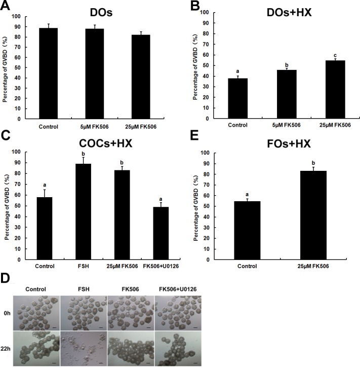
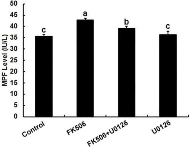
Calcineurin (CN) is a serine/threonine phosphatase which plays important roles in meiosis maturation in invertebrate oocytes; however, the role of CN in mouse oocytes is relatively unexplored. In this study, we examined the expression, localization and functional roles of CN in mouse oocytes and granulosa cells. The RT-PCR results showed that the β isoform of calcineurin A subunit (Cn A) expressed significantly higher than α and γ isoforms, and the expression of Cn Aβ mRNA obviously decreased in oocytes in which germinal vesicle breakdown (GVBD) occurred, while only B1 of calcineurin B subunit (Cn B) was detected in oocytes and stably expressed during oocytes maturation. The following fluorescence experiment showed that Cn A was mainly located in the nucleus of germinal vesicle (GV) stage oocytes and gruanlosa cells, and subsequently dispersed into the entire cytoplasm after GVBD. The decline of Cn A in oocytes suggested that it may play an important role in GVBD. To further clarify the role of calcineurin during meiotic maturation, FK506 (a calcineurin inhibitor) was used in the culture medium contained hypoxanthine (HX) which could keep mouse oocytes staying at GV stage. As expected, FK506 could induce a significant elevation of GVBD rate and increase the MPF level of denuded oocytes (DOs). Furthermore, FK506 could also play an induction role of GVBD of oocytes in COCs and follicles, and the process could be counteracted by MAPK kinase inhibitor (U0126). Above all, the results implied that calcineurin might play a crucial role in development of mouse oocytes and MPF and MAPK pathways are involved in this process.
Keywords: Calcineurin; FK506; GVBD; MAPK; MPF.
Conflict of interest statement
The authors declare there are no competing interests.
Figures
Similar articles
-
Activities of maturation-promoting factor (MPF) and mitogen-activated protein kinase (MAPK) are not required for the global histone deacetylation observed after germinal vesicle breakdown (GVBD) in porcine oocytes.Reproduction. 2006 Mar;131(3):439-47. doi: 10.1530/rep.1.00924. Reproduction. 2006. PMID: 16514187
-
Resumption of meiosis induced by meiosis-activating sterol has a different signal transduction pathway than spontaneous resumption of meiosis in denuded mouse oocytes cultured in vitro.Biol Reprod. 2001 Dec;65(6):1751-8. doi: 10.1095/biolreprod65.6.1751. Biol Reprod. 2001. PMID: 11717137
-
Mitogen-activated protein kinase translocates into the germinal vesicle and induces germinal vesicle breakdown in porcine oocytes.Biol Reprod. 1998 Jan;58(1):130-6. doi: 10.1095/biolreprod58.1.130. Biol Reprod. 1998. PMID: 9472933
-
The effect of hypoxanthine on mouse oocyte growth and development in vitro: maintenance of meiotic arrest and gonadotropin-induced oocyte maturation.Dev Biol. 1987 Feb;119(2):313-21. doi: 10.1016/0012-1606(87)90037-6. Dev Biol. 1987. PMID: 3100361
-
Morphological dynamics of cumulus-oocyte complex during oocyte maturation.Ital J Anat Embryol. 1998;103(4 Suppl 1):103-18. Ital J Anat Embryol. 1998. PMID: 11315942 Review.
Cited by
-
Proteomic Exploration of Porcine Oocytes During Meiotic Maturation in vitro Using an Accurate TMT-Based Quantitative Approach.Front Vet Sci. 2022 Feb 7;8:792869. doi: 10.3389/fvets.2021.792869. eCollection 2021. Front Vet Sci. 2022. PMID: 35198619 Free PMC article.
-
Increased transcriptome variation and localised DNA methylation changes in oocytes from aged mice revealed by parallel single-cell analysis.Aging Cell. 2020 Dec;19(12):e13278. doi: 10.1111/acel.13278. Epub 2020 Nov 17. Aging Cell. 2020. PMID: 33201571 Free PMC article.
References
-
- Akool El S, Gauer S, Osman B, Doller A, Schulz S, Geiger H, Pfeilschifter J, Eberhardt W. Cyclosporin A and tacrolimus induce renal Erk1/2 pathway via ROS-induced and metalloproteinase-dependent EGF-receptor signaling. Biochemical Pharmacology. 2012;83:286–295. doi: 10.1016/j.bcp.2011.11.001. - DOI - PubMed
-
- Bandyopadhyay J, Lee J, Lee J, Lee JI, Yu JR, Jee C, Cho JH, Jung S, Lee MH, Zannoni S, Singson A, Kim DH, Koo HS, Ahnn J. Calcineurin, a calcium/calmodulin-dependent protein phosphatase, is involved in movement, fertility, egg laying, and growth in Caenorhabditis elegans. Molecular Biology of the Cell. 2002;13:3281–3293. doi: 10.1091/mbc.E02-01-0005. - DOI - PMC - PubMed
-
- Choi T, Rulong S, Resau J, Fukasawa K, Matten W, Kuriyama R, Mansour S, Ahn N, Vande Woude GF. Mos/mitogen-activated protein kinase can induce early meiotic phenotypes in the absence of maturation-promoting factor: a novel system for analyzing spindle formation during meiosis I. Proceedings of the National Academy of Sciences of the United States of America. 1996;93:4730–4735. doi: 10.1073/pnas.93.10.4730. - DOI - PMC - PubMed
LinkOut - more resources
Full Text Sources
Other Literature Sources
Research Materials
Miscellaneous
