Review
. 2017 Jul;74(13):2413-2438.
doi: 10.1007/s00018-017-2478-0. Epub 2017 Feb 27.

Deciphering the BAR code of membrane modulators

Affiliations
Review

Deciphering the BAR code of membrane modulators

Ulrich Salzer et al. Cell Mol Life Sci. 2017 Jul.

Abstract

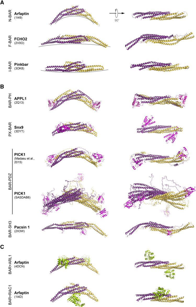
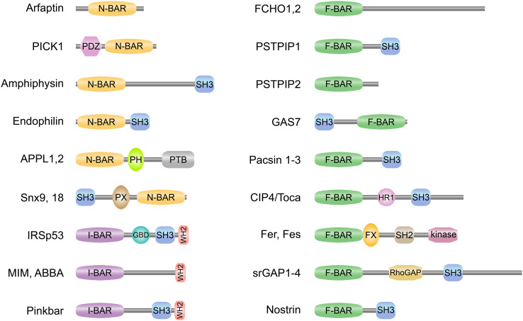
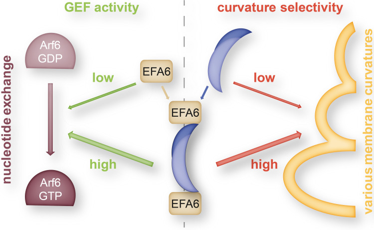
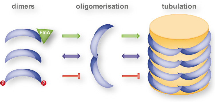
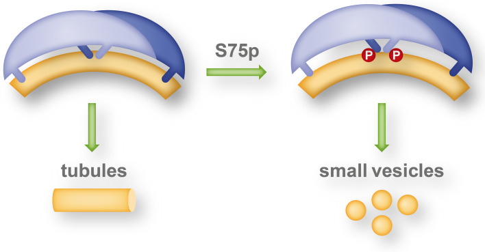
The BAR domain is the eponymous domain of the "BAR-domain protein superfamily", a large and diverse set of mostly multi-domain proteins that play eminent roles at the membrane cytoskeleton interface. BAR domain homodimers are the functional units that peripherally associate with lipid membranes and are involved in membrane sculpting activities. Differences in their intrinsic curvatures and lipid-binding properties account for a large variety in membrane modulating properties. Membrane activities of BAR domains are further modified and regulated by intramolecular or inter-subunit domains, by intermolecular protein interactions, and by posttranslational modifications. Rather than providing detailed cell biological information on single members of this superfamily, this review focuses on biochemical, biophysical, and structural aspects and on recent findings that paradigmatically promote our understanding of processes driven and modulated by BAR domains.

Keywords: F-BAR domain; I-BAR domain; Membrane curvature; Membrane remodelling; N-BAR domain; lipid binding.


Conflict of interest statement

The authors declare no conflict of interest.

Figures

Fig. 1
Fig. 1
Structure of selected BAR domain dimers. The BAR domain dimers form an elongated structure with a core bundle of six α-helices generated by antiparallel dimerisation of two BAR domain monomers. 3D structures of BAR domain dimers are shown as a ribbon. Monomers are depicted in different colors (yellow and dark magenta). Side view of the each BAR dimer is shown on left, while top view is on right. a Examples of BAR domain dimers representing N-BAR, F-BAR, and I-BAR domain fold. Different degrees of curvature adopted by each class of BAR domain dimers are depicted by grey lines. b Structures of BAR domain dimers from different subfamilies with their accessory domains (PH, PX, PDZ, and SH3) shown in magenta. Note, for PICK1, that two SAXS analysis derived models are shown. In PICK1 model (SASDAB8), the PDZ domains are far apart and flexible with respect to the BAR domain. Here, overlay of three generated models is shown. In the PICK1 model of Madasu et al. [78], the position of the PDZ domain was found to be well constrained, and packed against BAR domain. c Structure of the Arfaptin-2 BAR domain dimer in complex with Arl1 GTPase, and Rac1-GDP, both shown in green
Fig. 2
Fig. 2
Schematic domain representation of selected BAR domain proteins. Selected members of the N-BAR (BAR with an N-terminal amphipathic helix) and I-BAR (inverse-BAR) domain family on left and F-BAR (Fes/CIP4 homology-BAR) domain family on right are depicted. Most BAR domain proteins contain one or several additional domains with lipid-binding, protein-binding, and/or enzymatic activities. PDZ (PSD95/Dlg1/ZO-1) domain mediates protein–protein interactions by binding to the C-terminus of other specific proteins. SH3 (Src homology 3) domain confers binding to poly-proline motifs of target proteins, like N-WASP or dynamin. The phosphoinositides-binding PX (phox homology) and PH (pleckstrin homology) domains modulate membrane-binding specificities of different subsets of N-BAR domain proteins. PTB (phosphotyrosine-binding) domain binds to phosphotyrosine. GBD (GTPase-binding domain) is required for binding to Rho small GTPases. WH2 (Wiskott-Aldrich syndrome homology 2) domain binds to actin monomers and can facilitate the assembly of actin monomers into actin filaments. HR1 (protein kinase C-related kinase homology region 1) binds the small G protein Rho. FX (F-BAR extension) domain in Fer was shown to bind phosphatidic acid. SH2 (Src homology 2) domain allows binding to phosphorylated tyrosine residues on other proteins. RhoGAP (Rho GTPase activating protein) domain modulates the activity of Rho. Fer and Fes possess a tyrosine kinase domain
Fig. 3
Fig. 3
Complex between endophilin and EFA6 as regulator in clathrin-mediated endocytosis. The N-BAR domain of the endophilin dimer (dark and light blue overlapping moons representing endophilin monomers) interacts with the Arf6-specific exchange factor (EFA6). The complex exhibits both increased guanine-nucleotide exchange factor (GEF) activity of the EFA6 constituents and increased selectivity of the endophilin dimer for highly curved membrane shapes (yellow) and thereby plays a crucial role in orchestrating the sequential steps (Arf6 activation and selective membrane tubulation) in clathrin-mediated endocytosis [101]
Fig. 4
Fig. 4
Model for pacsin-2 bound to F-actin, based on EM reconstruction of F-actin decorated by pacin-2 [50]. Actin subunits (magenta ribbons) are numbered along one strand. The two green pacsin-2 ribbons on the right bind to that strand. The green pacsin-2 ribbon on the left binds to the opposite actin strand. The yellow surface at the bottom is a three-dimensional reconstruction of the atomic model shown, after imposing the actin helical symmetry and filtering to 12 Å resolution. Residues of the wedge loop, pointing towards F-actin, are represented as blue spheres
Fig. 5
Fig. 5
Regulation of pacsin’s membrane activity. Inter-dimer tip-to-tip oligomerization and membrane tubulation are reversible processes dependent on the pacsin dimer (dark and light blue moons) concentration. Phosphorylation at T181 (red circles) located at the tips of the dimers inhibits [137], whereas the presence of filamin A (FlnA) promotes [114] oligomerisation and pacsin-dependent membrane tubulation
Fig. 6
Fig. 6
Phosphorylation of the central insert of endophilin A1 controls its generation of membrane shapes. Apart from its N-terminal amphipathic helices at the tips of the BAR-domain dimer, endophilin A1 (dark and light blue moons) contains an additional pair of amphipathic helices (also termed central insert region). Shallow insertion of the amphipathic helices preferentially stabilizes small vesicles, whereas deep insert of these helices and tight contact of the BAR-domain with the headgroups of the membrane phospholipids favor membrane tubulation. Phosphorylation of the central insert region at S75 (red circles) by the Parkinson disease-associated kinase LRKK2 controls the membrane insertion depth of the amphipathic helices and thereby the type of membrane curvature generated by endophilin A1 [58]

References

    1. Sakamuro D, Elliott KJ, Wechsler-Reya R, Prendergast GC. BIN1 is a novel MYC-interacting protein with features of a tumour suppressor. Nat Genet. 1996;14(1):69–77. doi: 10.1038/ng0996-69. - DOI - PubMed
    1. Peter BJ, Kent HM, Mills IG, Vallis Y, Butler PJ, Evans PR, McMahon HT. BAR domains as sensors of membrane curvature: the amphiphysin BAR structure. Science. 2004;303(5657):495–499. doi: 10.1126/science.1092586. - DOI - PubMed
    1. Itoh T, Erdmann KS, Roux A, Habermann B, Werner H, De Camilli P. Dynamin and the actin cytoskeleton cooperatively regulate plasma membrane invagination by BAR and F-BAR proteins. Dev Cell. 2005;9(6):791–804. doi: 10.1016/j.devcel.2005.11.005. - DOI - PubMed
    1. Tsujita K, Suetsugu S, Sasaki N, Furutani M, Oikawa T, Takenawa T. Coordination between the actin cytoskeleton and membrane deformation by a novel membrane tubulation domain of PCH proteins is involved in endocytosis. J Cell Biol. 2006;172(2):269–279. doi: 10.1083/jcb.200508091. - DOI - PMC - PubMed
    1. Suetsugu S, Murayama K, Sakamoto A, Hanawa-Suetsugu K, Seto A, Oikawa T, Mishima C, Shirouzu M, Takenawa T, Yokoyama S. The RAC binding domain/IRSp53-MIM homology domain of IRSp53 induces RAC-dependent membrane deformation. J Biol Chem. 2006;281(46):35347–35358. doi: 10.1074/jbc.M606814200. - DOI - PubMed

Publication types

Substances

LinkOut - more resources