Review
. 2017 Feb 24;6(1):17.
doi: 10.3390/biology6010017.

Glutamine-Glutamate Cycle Flux Is Similar in Cultured Astrocytes and Brain and Both Glutamate Production and Oxidation Are Mainly Catalyzed by Aspartate Aminotransferase

Affiliations
Review

Glutamine-Glutamate Cycle Flux Is Similar in Cultured Astrocytes and Brain and Both Glutamate Production and Oxidation Are Mainly Catalyzed by Aspartate Aminotransferase

Leif Hertz et al. Biology (Basel). .

Abstract

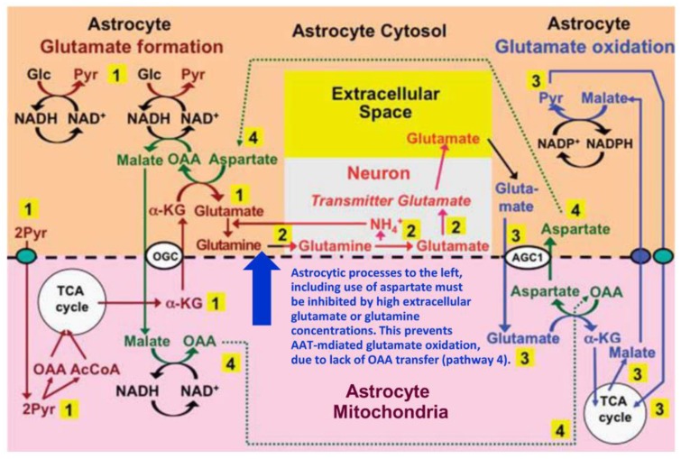
The glutamine-glutamate cycle provides neurons with astrocyte-generated glutamate/γ-aminobutyric acid (GABA) and oxidizes glutamate in astrocytes, and it returns released transmitter glutamate/GABA to neurons after astrocytic uptake. This review deals primarily with the glutamate/GABA generation/oxidation, although it also shows similarity between metabolic rates in cultured astrocytes and intact brain. A key point is identification of the enzyme(s) converting astrocytic α-ketoglutarate to glutamate and vice versa. Most experiments in cultured astrocytes, including those by one of us, suggest that glutamate formation is catalyzed by aspartate aminotransferase (AAT) and its degradation by glutamate dehydrogenase (GDH). Strongly supported by results shown in Table 1 we now propose that both reactions are primarily catalyzed by AAT. This is possible because the formation occurs in the cytosol and the degradation in mitochondria and they are temporally separate. High glutamate/glutamine concentrations abolish the need for glutamate production from α-ketoglutarate and due to metabolic coupling between glutamate synthesis and oxidation these high concentrations render AAT-mediated glutamate oxidation impossible. This necessitates the use of GDH under these conditions, shown by insensitivity of the oxidation to the transamination inhibitor aminooxyacetic acid (AOAA). Experiments using lower glutamate/glutamine concentration show inhibition of glutamate oxidation by AOAA, consistent with the coupled transamination reactions described here.

Keywords: aspartate aminotransferase; astrocyte culture; brain metabolism; glutamate dehydrogenase; glutamate oxidation; glutamate-glutamine cycle; glutamine synthetase; malate-aspartate shuttle; metabolic compartmentation; nitrogen balance.


Conflict of interest statement

The authors declare no conflict of interest.

Figures

Figure 3
Figure 3
Proposed pathway for coupled production and metabolism of transmitter glutamate using aspartate transamination for exchange between α-KG and glutamate. Joint pyruvate carboxylase and pyruvate dehydrogenase activation generates a “new” molecule of citrate (lower left corner) as detailed in Figure 1. Citrate-derived α-KG exiting the mitochondrial membrane leaves the astrocytic TCA cycle and is transaminated with aspartate to form glutamate, with concomitant oxaloacetate (OAA) formation from aspartate. The mitochondrial exit of α-KG occurs via the α-ketoglutarate/malate exchanger, generally acknowledged to be expressed in astrocytes, and the cytosolic malate with which it is exchanged, is generated via NADH-supported reduction of oxaloacetate generated from aspartate. Glutamate is amidated to glutamine (pathway 1), which is transferred to glutamatergic neurons (without indication of any extracellular space in the Figure). High extracellular concentrations of glutamate or glutamine (blue arrow) will at least in cultured astrocytes make the astrocytic production of glutamate and glutamine unnecessary (and/or prevent the reaction for thermodynamic reasons) and thereby inhibit glutamate formation from α-KG and the associated transamination of aspartate. In neurons glutamine is in a complex pathway converted to glutamate, accumulated in vesicles and released as transmitter glutamate (pathway 2). Subsequent reuptake of glutamate and oxidative metabolism in astrocytes (pathway 3) is normally of similar magnitude as the production of glutamate described above. Cytosolic glutamate is transferred to mitochondria via the aspartate-glutamate exchanger AGC1 (aralar) in exchange with mitochondrial aspartate generated from OAA formed during the synthesis of glutamate (pathway 4) and re-converted to aspartate during transamination of glutamate to α-KG. In turn, the cytosolic aspartate is used during glutamate synthesis in the transamination of α-KG to glutamate in pathway 1 after transfer via pathway 4. However, when glutamate production from α-KG is inhibited by high extracellular concentrations of glutamate or glutamine (blue arrow) or by excess ammonia (inhibiting the neuronal glutaminase [98]), the exit of aspartate to the cytosol in exchange with glutamate during glutamate oxidation will no longer be compensated for by aspartate use during glutamate formation. This abolishes glutamate oxidation via AAT, so that formation of α-KG must be catalyzed by glutamate dehydrogenase (GDH). This is unlikely normally to take place in vivo since elevated extracellular glutamate only occurs during bursts of neuronal activity and glutamate uptake and glutamine synthesis rapidly clear extra- and intracellular glutamate. Metabolism of α-KG is only shown via malate exit and pyruvate formation. Since this is an oxidation in the cytosol it must be followed by malate aspartate shuttle (MAS)-mediated transfer of a reducing equivalent to the mitochondria. Biosynthesis of glutamine is shown in brown and metabolic degradation of glutamate in blue. Synthesis of glutamine and export from the glia is critical since it restores nitrogen balance (otherwise concentrations of glutamate and aspartate would continuously rise). Redox shuttling and astrocytic release of glutamine and uptake of glutamate are shown in black, and neuronal uptake of glutamine, hydrolysis to glutamate, and its release is shown in red. Reactions involving or resulting from transamination between aspartate and oxaloacetate (OAA) are shown in green. Small blue oval shows pyruvate carrier into mitochondria and small purple oval malate carrier out from mitochondria. AGC1, aspartate/glutamate exchanger, aralar; α-KG, α-ketoglutarate; Glc, glucose; Pyr, pyruvate; OGC, malate/α-ketoglutarate exchanger. AGC1 is for graphical reasons only indicated during the initial part of glutamate oxidation, where it constitutes part of the suggested pathway, but not where it is generally acknowledged to function in MAS, i.e., during synthesis of pyruvate from glucose (pathway 1) and from malate (pathway 3). The glutamine–(GABA) cycle is accordingly extremely dependent upon the abundant expression of AGC1 in astrocytes described above. However, if malate generated during glutamate degradation does not exit the TCA cycle but is further metabolized to α-KG, allowing re-synthesis of another molecule of glutamate from only one molecule of pyruvate and abrogating pyruvate formation from malate (Figure 1) trafficking via AGC1 will be considerably reduced, although certainly not abolished. Slightly modified from [15].
Figure 1
Figure 1
Cartoon of glucose metabolism via pyruvate in neurons (left—N) and astrocytes (right—A) and of glutamine-glutamate (γ-aminobutyric acid, GABA) cycling. This figure shows (i) metabolic pathways; (ii) metabolic rates (µmol/min per 100 mg protein or 1 g wet wt); and (iii) inhibition by excess extracellular glutamate or glutamine. One molecule of glucose is metabolized by glycolysis in the cytosol to two molecules of pyruvate in a complex and strictly regulated pathway (not shown). In both neurons and astrocytes pyruvate metabolism via acetyl coenzyme A (ac.CoA) leads to formation of citrate in the tricarboxylic acid (TCA) cycle by condensation with preexisting oxaloacetate (OAA), an end result of the previous turn of the cycle. Citrate oxidation in the TCA cycle includes two decarboxylations, resulting in re-formation of oxaloacetate, ready for another turn of the cycle, and reduction of NAD+ to NADH (and a single FAD to FADH2), leading to large amounts of energy (ATP) via re-oxidation in the electron transport chain. Pyruvate carboxylation, which is active in astrocytes, but not in neurons, creates a new molecule of oxaloacetate, which after condensation with acetyl coenzyme A, forms citrate that is metabolized in the TCA cycle to α-ketoglutarate (α-KG), which can leave the cycle to form glutamate (glu), catalyzed by aspartate aminotransferase (AAT). Further metabolism by the cytosolic and astrocyte-specific enzyme glutamine synthetase leads to the formation of glutamine (gln), which after transport to neurons is converted to transmitter glutamate or GABA in complex reactions (reviewed in [6]). Released transmitter glutamate is almost quantitatively re-accumulated in astrocytes, together with at least part of the released GABA (upper line of glu-gln cycle) and re-accumulated in the astrocytic cytosol. Here, 75%–80% is converted to glutamine and re-enters the glutamine-glutamate (GABA) cycle. The remaining 20%–25% is oxidatively degraded. This paper suggests that the default mechanism for the initial conversion of glutamate to α-KG is also transamination by AAT (see Figure 3), but it does not exclude a minor contribution by glutamate dehydrogenase (GDH). Operation of AAT in two opposite directions is thermodynamically possible since the reactions take place in two different compartments (cytosol and mitochondria) and also are temporally separate. α-KG is metabolized via malate, which can exit to the cytosol and be decarboxylated by cytosolic malic enzyme to pyruvate, which is oxidized in the TCA cycle via acetyl coenzyme A. Another possibility is that malate does not exit the TCA cycle but is further metabolized to α-KG after condensation with acetyl coenzyme A, allowing re-synthesis of another molecule of glutamate from only one molecule of pyruvate [6]. The degraded glutamate/GABA must in the long term be replaced by a quantitatively similar production of glutamate from glucose, in the first case by complete de novo synthesis from one molecule glucose, in the second from one half of a glucose molecule. However, temporary fluctuations in the content of glutamate occur. The initial part of GABA metabolism is different, as all GABA is metabolized via succinic semialdehyde, succinate and α-KG to glutamate. Numbers in black show rates re-calculated as µmol/min per 100 mg protein based on rate of glucose uptake in our own cultured astrocytes, and those in red are in vivo rates from [10]. The percentage distribution between metabolism via pyruvate carboxylation and acetyl coenzyme A is based on averages of results tabulated in [12]. Those for glutamate metabolism to either α-KG or glutamine are from [10]. The in vivo results that 40% of 0.8 µmol/min per 100 mg protein is metabolized via pyruvate carboxylation is consistent with the pyruvate carboxylation rates found by Kaufman and Driscoll [13] and overall the correlation between the metabolic rates in vivo and in culture is remarkably good, although other authors have described lower rates of glucose metabolism in cultured astrocytes. It should also be kept in mind that lightly anaesthetized mice have been used in most in vivo studies. Blue arrows: Suggested prevention of glutamate formation from α-KG and accompanying aspartate utilization by the presence of high extracellular concentrations of glutamate or glutamine, with the consequence that the AAT-mediated, coupled glutamate formation and degradation illustrated in Figure 3 can no longer operate. This appears to be the reason that so many studies have concluded that glutamate oxidation is catalyzed by GDH, whereas studies using low glutamate/glutamine concentrations find that AAT is involved. Modified from [6].
Figure 2
Figure 2
Aspartate promotes glutamate synthesis in cultured astrocytes, but only in the absence of extracellular glutamate. (A,B) Cortical mouse astrocyte cultures were incubated for 1 h in saline (in mM: 140 NaCl, 3.6 KCl, 0.5 NaH2PO4, 0.5 MgSO4, 1.5 CaCl2, 2 NaHCO3, 10 HEPES, pH 7.4) containing 2 mM glucose in the absence or presence of added amino acids (γ-aminobutyric acid, GABA, aspartate, alanine, or leucine; 10–200 μmol/L). Glutamate (A) and glutamine (B) contents were measured in cellular extracts and media by enzymatic methods. (C,D) Similar measurements in the absence of added amino acid (Ctr), the presence of 50 μM aspartate (Asp); 50 μM glutamate (Glu) or both together (Asp/Glu). Note that aspartate is the only amino acid among those studied which increases glutamate (A) and glutamine (B) and that the effect is abolished in the presence of glutamate (C/D). From [78].

Similar articles

Cited by

References

    1. Smith Q.R. Transport of glutamate and other amino acids at the blood-brain barrier. J. Nutr. 2000;130:1016S–1022S. - PubMed
    1. Patel A.B., de Graaf R.A., Rothman D.L., Behar K.L. Effects of gamma-aminobutyric acid transporter 1 inhibition by tiagabine on brain glutamate and gamma-aminobutyric acid metabolism in the anesthetized rat in vivo. J. Neurosci. Res. 2015;93:1101–1108. doi: 10.1002/jnr.23548. - DOI - PMC - PubMed
    1. Danbolt N.C., Furness D.N., Zhou Y. Neuronal vs glial glutamate uptake: Resolving the conundrum. Neurochem. Int. 2016;98:29–45. doi: 10.1016/j.neuint.2016.05.009. - DOI - PubMed
    1. Duarte J.M., Gruetter R. Glutamatergic and gabaergic energy metabolism measured in the rat brain by 13C NMR spectroscopy at 14.1 T. J. Neurochem. 2013;126:579–590. doi: 10.1111/jnc.12333. - DOI - PubMed
    1. Schousboe A., Bak L.K., Waagepetersen H.S. Astrocytic control of biosynthesis and turnover of the neurotransmitters glutamate and GABA. Front Endocrinol (Lausanne) 2013;4 doi: 10.3389/fendo.2013.00102. - DOI - PMC - PubMed

LinkOut - more resources