. 2017 Feb 24;18(3):487.
doi: 10.3390/ijms18030487.

Pancreatic Cancer-Induced Neutrophil Extracellular Traps: A Potential Contributor to Cancer-Associated Thrombosis

Affiliations

Pancreatic Cancer-Induced Neutrophil Extracellular Traps: A Potential Contributor to Cancer-Associated Thrombosis

Norbaini Abdol Razak et al. Int J Mol Sci. .

Abstract

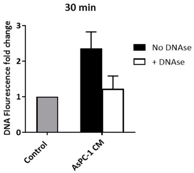
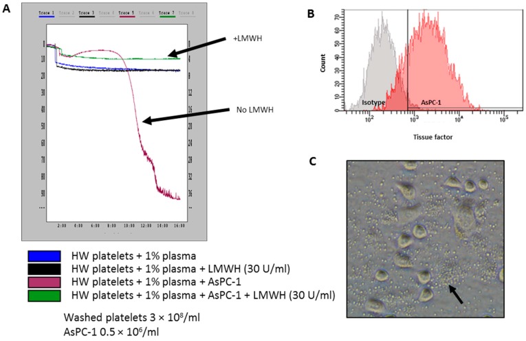
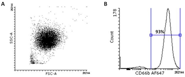
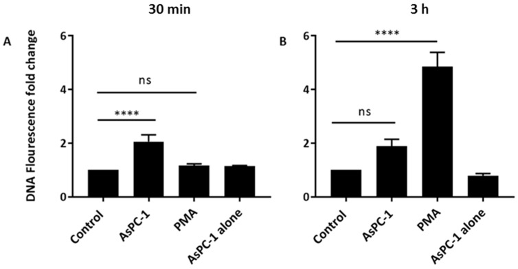
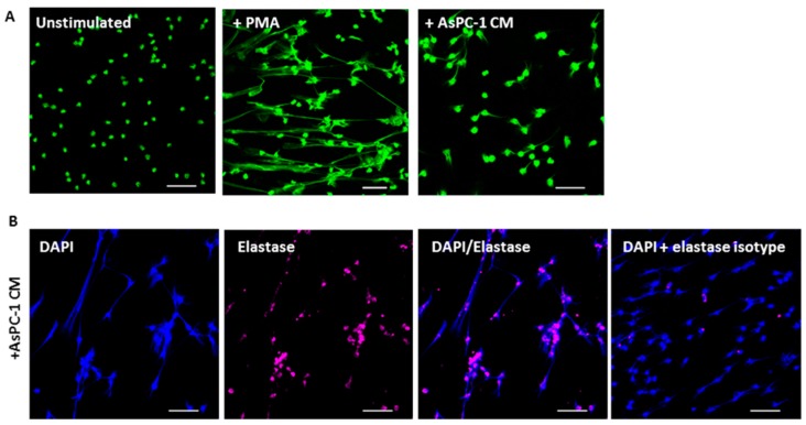
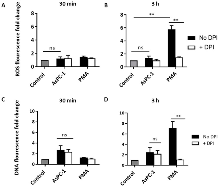
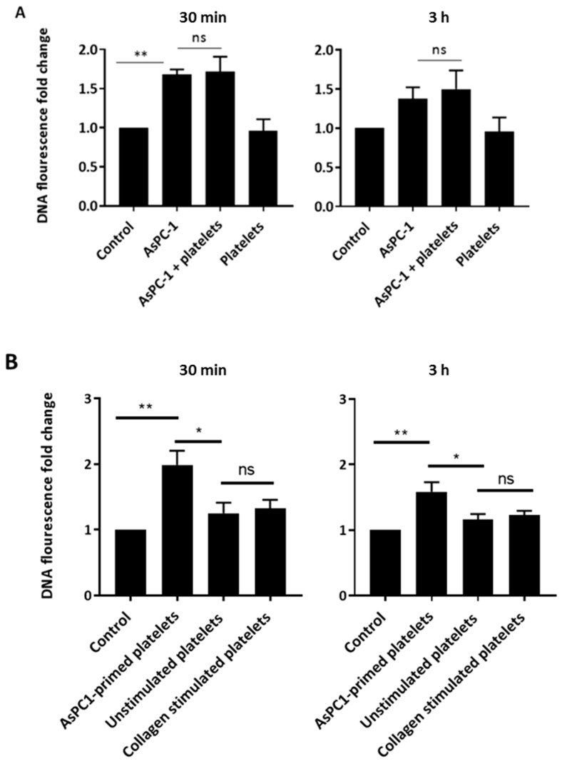
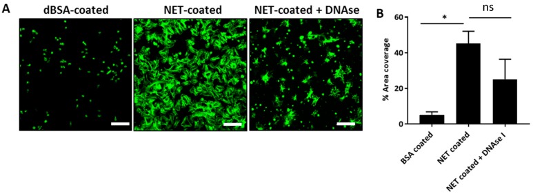
Pancreatic cancer (PaCa) is a highly metastatic cancer, and patients are at high risk of developing venous thromboembolism (VTE). Neutrophil extracellular traps (NETs) have been associated with cancer metastasis and cancer-associated thrombosis, but the ability of cancer to stimulate NET release is not known. The release of NETs has been shown to be a slow process and requires reactive oxygen species (ROS) production. Studies suggest that activated platelets are important mediators in the release. Here, we show that PaCa cells can stimulate the rapid release of NETs, independently of ROS production. We further assessed the role of platelets in PaCa-induced NETs and observed a trend of increased the NET release by PaCa-primed platelets. Additionally, NETs promoted thrombus formation under venous shear stress ex vivo. Taken together, our results suggest that PaCa-induced NETs can contribute to the high risk of venous thromboembolism development in PaCa patients, and reveal NETs as a potential therapeutic target.

Keywords: neutrophil extracellular traps; pancreatic cancer; platelets; venous thromboembolism.


Conflict of interest statement

The authors declare no conflict of interest.

Figures

Figure A1
Figure A1
The addition of DNAse I (60 U/mL) abolished extracellular DNA fluorescence to unstimulated control, confirming the specificity of Sytox Green to extracellular DNA (exDNA). Control: unstimulated neutrophils. Data expressed as mean ± SEM from n = 3.
Figure A2
Figure A2
AsPC-1 expresses tissue factor, and causes platelet aggregation (A) Light transmission aggregometry showed that AsPC-1-induced aggregation of human washed (HW) platelets, which was tissue factor-dependent, as addition of low-molecular-weight heparin (LMWH) prevented platelet aggregation. The downward-slope represents light transmission approaching 100%, which is indicative of platelet aggregation; (B) Flow cytometry analysis of AsPC-1 cells stained with anti-tissue factor show AsPC-1 expressing tissue factor above isotype control; (C) Phase-contrast images show platelets aggregating (black arrow) around AsPC-1 cells.
Figure A3
Figure A3
Flow cytometry analysis of CD66b staining on isolated human neutrophils. (A) Forward-side-scatter profile of isolated polymorphonuclear neutrophils (PMNs); (B) Isolated PMNs were stained with CD66b, a marker for PMNs, to determine purity of neutrophil isolation. A total of 93% of the isolated cell population was positive for CD66b.
Figure 1
Figure 1
AsPC-1 stimulates the rapid release of neutrophil extracellular traps (NETs) from human neutrophils. Human neutrophils were incubated with AsPC-1 cells at a neutrophil to AsPC-1 ratio of 1:2 and NETs were quantified using Sytox Green (5 μM) in a fluorescence plate reader at (A) 30 min and (B) 3 h. AsPC-1 induced rapid NET release at 30 min while phorbyl myristate acetate (PMA)-stimulated neutrophils (positive control) required 3 h. Control: unstimulated neutrophils. (n = 12, **** p < 0.0001 and ns = non-significant); One-way ANOVA followed by Bonferroni post-test. Data presented as mean ± SEM.
Figure 2
Figure 2
AsPC-1 stimulates NET release via soluble protein mediators. (A) The conditioned media (CM) of AsPC-1 cells stimulated the release of NETs. Mesenchymal stem cell (MSC) CM, which served as a control for human non-malignant cells, did not stimulate NET release (n = 3, * p < 0.01 and ns = non-significant.); (B) AsPC-1 CM was separated into lipid and protein fractions, and each was incubated with neutrophils for 30 min. The protein fraction of AsPC-1 CM stimulated NET release while the lipid fraction did not. Media RPMI fractions served as control (n = 3). Control: unstimulated neutrophils. One-way ANOVA followed by Bonferroni post-test. Data presented as mean ± SEM.
Figure 3
Figure 3
Confocal microscopy images of NETs induced by AsPC-1 conditioned media. (A) Neutrophils were incubated on poly-l-lysine coated glass slides and left either unstimulated, or were stimulated with PMA for 3 h, or AsPC-1 CM for 30 min. Neutrophils were fixed with paraformaldehyde and stained with Sytox (green) to visualise extracellular DNA. Images representative of three independent experiments; (B) AsPC-1 stimulated neutrophils were also stained with anti-elastase (pink) to visualise elastase bound on NETs. DAPI (blue) was also used as an alternative to visualise DNA. Elastase staining was above isotype control. Images representative of two independent experiments. Scale bar = 50 μm.
Figure 4
Figure 4
AsPC-1-induced-NET generation is independent of reactive oxygen species production. (A,B) Quantification of reactive oxygen species (ROS) generated during incubation with AsPC-1 CM and PMA at (A) 30 min and (B) 3 h using 2′,7′-dichlorodihydrofluorescein diacetate (H2DCFDA) fluorescent dye. ROS were not significantly generated in AsPC-1 CM-stimulated neutrophils at either time points; (B) PMA-stimulated neutrophils had significant ROS generated at 3 h, this was abolished with the addition of non-specific ROS inhibitor diphenyleneiodonium (DPI); (C,D) The effect of DPI on NET release in AsPC-1 or PMA-induced NETs; (C) DPI did not reduce AsPC-1-induced NETs; (D) NETs were significantly reduced in PMA-stimulated neutrophils by DPI. n = 3, ** p < 0.001, ns = non-significant. One-way ANOVA followed by Bonferroni post-test. Data presented as mean ± SEM.
Figure 5
Figure 5
AsPC-1-primed platelets, but not washed platelets, stimulate rapid release of NETs. (A) Platelets (1 × 10⁸/mL) were incubated with neutrophils and AsPC-1 cells. Platelets had no effect on AsPC-1-induced NETS; (B) the effect of AsPC-1-primed platelets on NET release. Platelets were primed with AsPC-1 cells by incubation with AsPC-1 for 4 h. The supernatant was removed and centrifuged to pellet platelets which were then resuspended and incubated with neutrophils. Primed platelets caused rapid NET release in 30 min. (n = 4, * p < 0.01, ** p < 0.001 and ns = non-significant. One-way ANOVA followed by Bonferroni post-hoc). Control: unstimulated neutrophils. Data expressed as mean ± SEM.
Figure 6
Figure 6
NETs promote static platelet adhesion and activation. (A) NETs were isolated from PMA-stimulated neutrophils before coated on glass slides. Washed platelets (2 × 10⁷/mL) were incubated with NETs, and platelet adhesion and spreading were assessed using confocal microscopy. Denatured bovine serum albumin (dBSA)-coated slides served as negative control. NETs caused platelets to adhere and spread. The pre-treatment with deoxyribonuclease I (DNAse I) did not completely abrogate adhesion and spreading. Images representative of three independent experiments. Scale bar = 20 μm; (B) quantification of percentage area coverage of platelet adhesion and spreading using Image J analysis software. n = 3,* p < 0.05 and ns = non-significant. One-way ANOVA followed by Bonferroni post-hoc. Data presented as mean ± SEM.
Figure 7
Figure 7
NETs entrap and activate platelets and promote thrombus formation. (A) 2D confocal images of platelet adhesion and thrombus formation in collagen-, dBSA-, and NET-coated channels after perfusing DiO6C(3) fluorescently-labelled whole blood for 10 min at 10 dyne/cm2. Platelet adhesion and thrombi are visualised in NET-coated channels. Collagen- and dBSA-coated channels served as positive and negative controls, respectively. Scale bar = 90 μm; (B) Z-stack images confirm the presence of thrombi on NETs, which are similar to those in collagen-coated channels; (C) Quantification of surface area coverage of platelet adhesion showed a significant increase in NET-coated channels. n = 3, * p < 0.05 unpaired t-test.

References

    1. Fuchs T.A., Abed U., Goosmann C., Hurwitz R., Schulze I., Wahn V., Weinrauch Y., Brinkmann V., Zychlinsky A. Novel cell death program leads to neutrophil extracellular traps. J. Cell Biol. 2007;176:231–241. doi: 10.1083/jcb.200606027. - DOI - PMC - PubMed
    1. Brinkmann V., Reichard U., Goosmann C., Fauler B., Uhlemann Y., Weiss D.S., Weinrauch Y., Zychlinsky A. Neutrophil extracellular traps kill bacteria. Science. 2004;303:1532–1535. doi: 10.1126/science.1092385. - DOI - PubMed
    1. Gupta A.K., Hasler P., Holzgreve W., Gebhardt S., Hahn S. Induction of neutrophil extracellular DNA lattices by placental microparticles and IL-8 and their presence in preeclampsia. Hum. Immunol. 2005;66:1146–1154. doi: 10.1016/j.humimm.2005.11.003. - DOI - PubMed
    1. Hakkim A., Fürnrohr B.G., Amann K., Laube B., Abed U.A., Brinkmann V., Herrmann M., Voll R.E., Zychlinsky A. Impairment of neutrophil extracellular trap degradation is associated with lupus nephritis. Proc. Natl. Acad. Sci. USA. 2010;107:9813–9818. doi: 10.1073/pnas.0909927107. - DOI - PMC - PubMed
    1. Yoo D.-G., Winn M., Pang L., Moskowitz S.M., Malech H.L., Leto T.L., Rada B. Release of cystic fibrosis airway inflammatory markers from pseudomonas aeruginosa-stimulated human neutrophils involves nadph oxidase-dependent extracellular DNA trap formation. J. Immunol. 2014;192:4728–4738. doi: 10.4049/jimmunol.1301589. - DOI - PMC - PubMed

Substances