Review
. 2017 Feb 25;18(3):504.
doi: 10.3390/ijms18030504.

The Diverse Roles of Microglia in the Neurodegenerative Aspects of Central Nervous System (CNS) Autoimmunity

Affiliations
Review

The Diverse Roles of Microglia in the Neurodegenerative Aspects of Central Nervous System (CNS) Autoimmunity

Kaitlyn K Thompson et al. Int J Mol Sci. .

Abstract

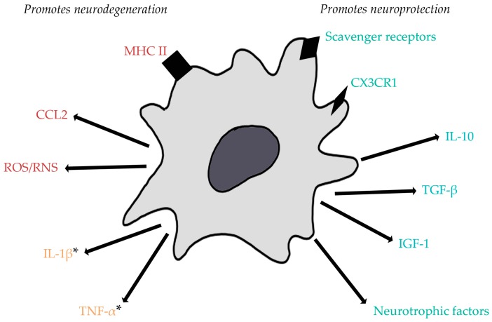
Autoimmune diseases of the central nervous system (CNS) involve inflammatory components and result in neurodegenerative processes. Microglia, the resident macrophages of the CNS, are the first responders after insults to the CNS and comprise a major link between the inflammation and neurodegeneration. Here, we will focus on the roles of microglia in two autoimmune diseases: the prevalent condition of multiple sclerosis (MS) and the much rarer Rasmussen's encephalitis (RE). Although there is an abundance of evidence that microglia actively contribute to neuronal damage in pathological states such as MS and RE, there is also evidence of important reparative functions. As current research supports a more complex and diverse array of functions and phenotypes that microglia can assume, it is an especially interesting time to examine what is known about both the damaging and restorative roles that microglia can play in the inflammatory CNS setting. We will also discuss the pharmacological approaches to modulating microglia towards a more neuroprotective state.

Keywords: Rasmussen’s encephalitis; autoimmunity; microglia; multiple sclerosis.


Conflict of interest statement

The authors declare no conflict of interest.

Figures

Figure 1
Figure 1
Electron micrograph of microglia. Activated microglia can contribute to neurodegeneration or neuroprotection (courtesy of Dr. Iordanis Gravanis).
Figure 2
Figure 2
Expression and release of factors by activated microglia can promote neurodegeneration or neuroprotection in situations of central nervous system (CNS) autoimmunity. Generally, pro-inflammatory cytokines and reactive oxygen/nitrogen species contribute to neuronal injury and CC chemokine ligand 2 (CCL2) can recruit immune cells that can be the cause of further neuronal injury. Additionally, upregulation of MHC II promotes efficient antigen presentation and can lead to autoreactive T cell activation. On the other hand, expression of scavenger receptors and the fractalkine receptor (CX3CR1) by microglia promotes phagocytic activity allowing for clearance of dead cells or myelin debris, as seen in multiple sclerosis (MS). Neurotrophic factors, anti-inflammatory cytokines, and insulin growth factor 1 (IGF-1) attenuate pro-inflammatory responses and promote tissue repair. * Though tumor necrosis factor α (TNF-α) has generally been considered more neurotoxic, low levels of this pro-inflammatory cytokine have recently been found to be neuroprotective in cases of excitotoxicity, which occurs in both MS and Rasmussen’s Encephalitis (RE). Both TNF-α and interleukin (IL)-1β have also been seen to be crucial for effective remyelination. ROS/RNS: reactive oxygen/nitrogen species. Red indicates factors that promote neurodegeneration; blue indicates factors that promote neuroprotection; yellow indicates factors shown to play both neurodegenerative and neuroprotective roles.

References

    1. Hewagama A., Richardson B. The genetics and epigenetics of autoimmune diseases. J. Autoimmun. 2009;33:3–11. doi: 10.1016/j.jaut.2009.03.007. - DOI - PMC - PubMed
    1. Nimmerjahn A., Kirchhoff F., Helmchen F. Resting microglial cells are highly dynamic surveillants of brain parenchyma in vivo. Science. 2005;308:1314–1318. doi: 10.1126/science.1110647. - DOI - PubMed
    1. Davalos D., Grutzendler J., Yang G., Kim J.V., Zuo Y., Jung S., Littman D.R., Dustin M.L., Gan W.-B. ATP mediates rapid microglial response to local brain injury in vivo. Nat. Neurosci. 2005;8:752–758. doi: 10.1038/nn1472. - DOI - PubMed
    1. Hanisch U.K., Kettenmann H. Microglia: Active sensor and versatile effector cells in the normal and pathologic brain. Nat. Neurosci. 2007;10:1387–1394. doi: 10.1038/nn1997. - DOI - PubMed
    1. Wolf S.A., Boddeke H.W., Kettenmann H. Microglia in physiology and disease. Annu. Rev. Physiol. 2016 doi: 10.1146/annurev-physiol-022516-034406. - DOI - PubMed

MeSH terms