

Hearing disorders in cats
- PMID: 28245737
- PMCID: PMC11119533
- DOI: 10.1177/1098612X17695062
Hearing disorders in cats
Abstract
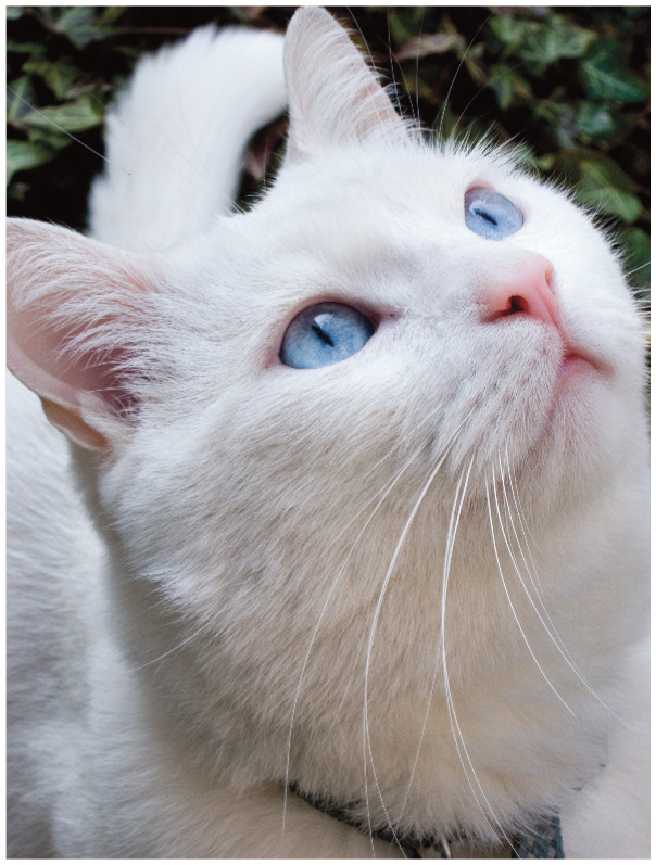
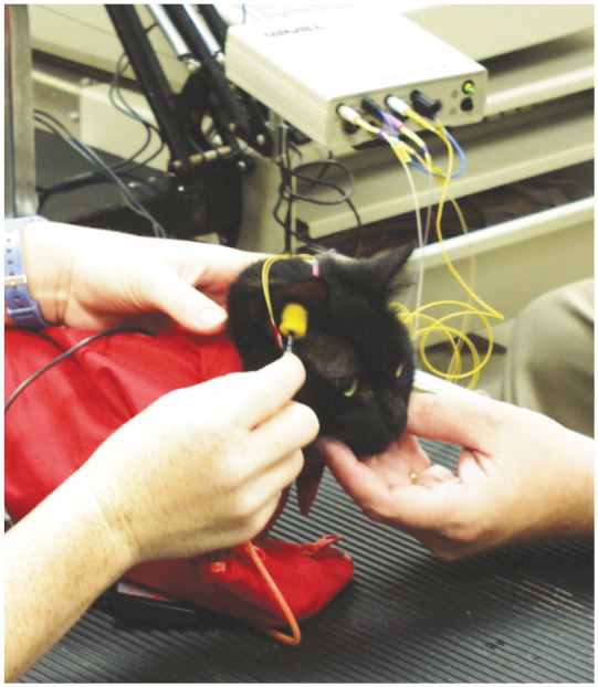
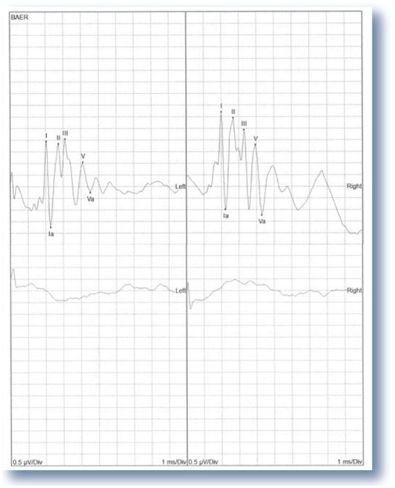
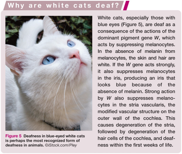
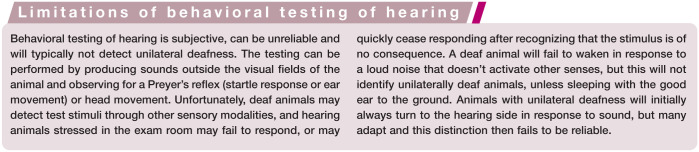
Practical relevance: Auditory function is a sense that is central to life for cats - being important in situational awareness of potential predators, pursuit of prey, and for communication with conspecifics, humans and other species. Deafness in cats is most frequently the result of a genetic disorder, strongly associated with white fur and blue eyes, but may also result from acquired causes such as advancing age, ototoxic drugs, infection, environmental noise and physical trauma. Deafness can be sensorineural, where there is loss of cochlear hair cells, or conductive, where sound is muffled on its way to the inner ear. Clinical challenges: Establishing whether a cat is deaf can be difficult as behavioral testing of hearing is subjective and does not reliably detect unilateral deafness. Brainstem auditory evoked response testing is an objective measure but is limited in its availability. Currently, sensorineural deafness is irreversible because no treatments are available to restore lost hair cells. Conductive hearing loss can usually be treated, although full hearing recovery following otitis media may take weeks as the body clears the middle ear of debris. Evidence base: The author draws on the published literature and his extensive research on clinical aspects and molecular genetics of deafness, principally in companion animals, to review types and forms of deafness in cats. He also discusses current diagnostic approaches and provides brief advice for managing cats with hearing loss.
Conflict of interest statement
The author declared no potential conflicts of interest with respect to the research, authorship, and/or publication of this article.
Figures
References
-
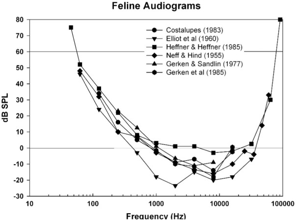
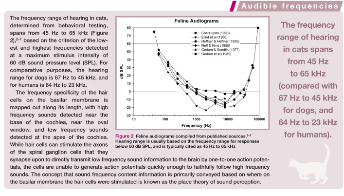
- Neff W, Hind J. Auditory thresholds of the cat. J Acoust Soc Am 1955; 27: 480-483.
-
- Elliot D, Stein L, Harrison M. Determination of absolute-intensity thresholds and frequency difference thresholds in cats. J Acoust Soc Am 1960; 32: 380-384.
-
- Gerken GM, Sandlin D. Auditory reaction time and absolute threshold in cat. J Acoust Soc Am 1977; 61: 602-607. - PubMed
-
- Costalupes JA. Temporal integration of pure tones in the cat. Hear Res 1983; 9: 43-54. - PubMed
Publication types
MeSH terms
LinkOut - more resources
Full Text Sources
Other Literature Sources
Medical
Miscellaneous
