. 2017 Feb 28;19(1):39.
doi: 10.1186/s13075-017-1237-9.

Epstein-Barr virus lytic infection promotes activation of Toll-like receptor 8 innate immune response in systemic sclerosis monocytes

Affiliations

Epstein-Barr virus lytic infection promotes activation of Toll-like receptor 8 innate immune response in systemic sclerosis monocytes

Antonella Farina et al. Arthritis Res Ther. .

Abstract

Background: Monocytes/macrophages are activated in several autoimmune diseases, including systemic sclerosis (scleroderma; SSc), with increased expression of interferon (IFN)-regulatory genes and inflammatory cytokines, suggesting dysregulation of the innate immune response in autoimmunity. In this study, we investigated whether the lytic form of Epstein-Barr virus (EBV) infection (infectious EBV) is present in scleroderma monocytes and contributes to their activation in SSc.

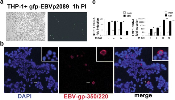
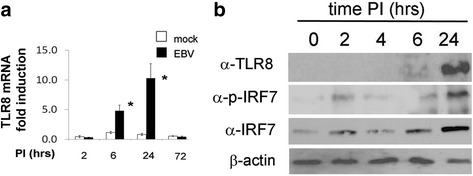
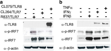
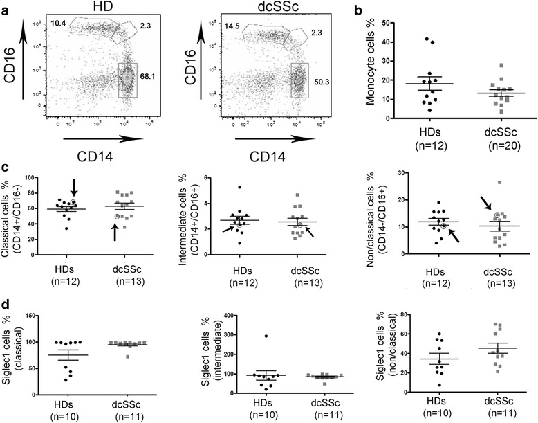
Methods: Monocytes were isolated from peripheral blood mononuclear cells (PBMCs) depleted of the CD19+ cell fraction, using CD14/CD16 negative-depletion. Circulating monocytes from SSc and healthy donors (HDs) were infected with EBV. Gene expression of innate immune mediators were evaluated in EBV-infected monocytes from SSc and HDs. Involvement of Toll-like receptor (TLR)8 in viral-mediated TLR8 response was investigated by comparing the TLR8 expression induced by infectious EBV to the expression stimulated by CL075/TLR8/agonist-ligand in the presence of TLR8 inhibitor in THP-1 cells.

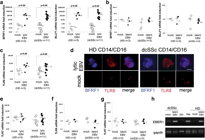
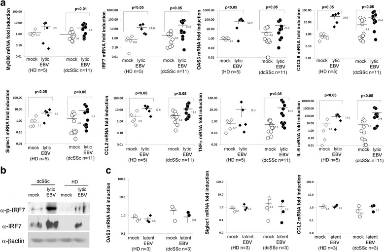
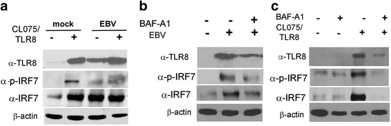
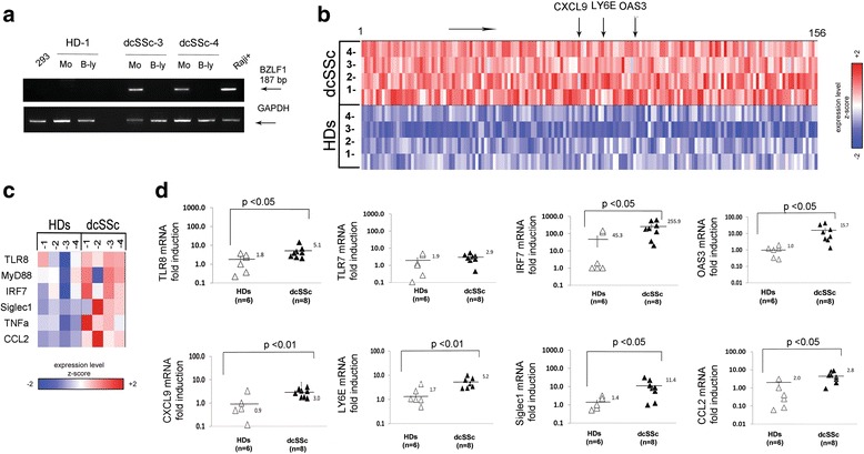
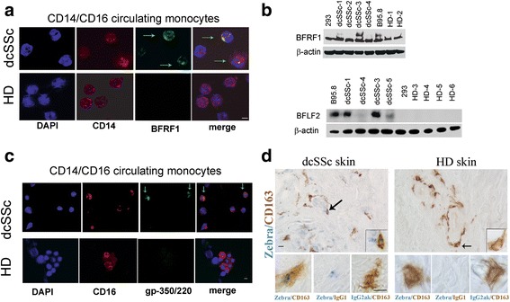
Results: Infectious EBV strongly induced TLR8 expression in infected SSc and HD monocytes in vitro. Markers of activated monocytes, such as IFN-regulated genes and chemokines, were upregulated in SSc- and HD-EBV-infected monocytes. Inhibiting TLR8 expression reduced virally induced TLR8 in THP-1 infected cells, demonstrating that innate immune activation by infectious EBV is partially dependent on TLR8. Viral mRNA and proteins were detected in freshly isolated SSc monocytes. Microarray analysis substantiated the evidence of an increased IFN signature and altered level of TLR8 expression in SSc monocytes carrying infectious EBV compared to HD monocytes.

Conclusion: This study provides the first evidence of infectious EBV in monocytes from patients with SSc and links EBV to the activation of TLR8 and IFN innate immune response in freshly isolated SSc monocytes. This study provides the first evidence of EBV replication activating the TLR8 molecular pathway in primary monocytes. Immunogenicity of infectious EBV suggests a novel mechanism mediating monocyte inflammation in SSc, by which EBV triggers the innate immune response in infected cells.

Keywords: EBV reactivation; IFN inducible genes; Innate immune response; Monocytes; Systemic sclerosis; Toll-like receptor 8.


Figures

Fig. 1
Fig. 1
EBV replication modulates TLR8 expression in monocytes in vitro. Negative selected monocytes from dcSSc patients and HDs were infected with EBV. For total RNA and immunostaining, monocytes were harvested 5 days PI. a,b,c,e,f,g mRNA expression of EBV-lytic and TLR genes was analyzed by quantitative PCR. Results are expressed as fold-change induction normalized by one of the mock infected healthy controls. 18S rRNA was used as internal control. Bars represent the mean ± SEM. The two-tailed t test was used for statistical analysis. d Immunofluorescence double staining shows EBV-BFRF1-antigen (blue) and TLR8 (red) co-expression in monocytes from one representative dcSSc and HD infected with EBV-p2089. Original magnification 60×, scale bar = 5 μm. h PCR products of EBV DNA in monocytes from representative dcSSc and HD; DNA from Raji-EBV-positive cells and DNA from 293 were used as positive control and negative control, respectively. GAPDH used as internal control. b, e, f and h refer to monocytes expressing EBV-p2089 latent infection. dcSSc diffuse cutaneous systemic sclerosis, EBV Epstein-Barr virus, HD healthy donor, TLR Toll-like receptor
Fig. 2
Fig. 2
EBV lytic genes modulate the expression of innate immune mediators, IFN-inducible genes, and proinflammatory cytokines in EBV-p2089-infected monocytes. a,c mRNA expression of indicated innate immune mediators and cytokines was evaluated by quantitative PCR in negative selected CD14/CD16 monocytes infected with EBV-p2089, 5 days PI. Results are expressed as the fold-change induction normalized by one of the mock infected healthy controls. 18S ribosomal RNA levels used as internal control. Bars represent the mean ± SEM. b Western blot analysis was performed to determine IRF7 protein (phosphorylated and total) levels in cell lysates of mock- and EBV-infected monocytes from one representative dcSSc patient and HD, 5 days PI. β-actin was used as loading control. Fold-changes shown on the graph are normalized to mRNA expression by one of the mock infected healthy controls. Bars represent mean ± S.E.M. The two-tailed t test was used for statistical analysis. dcSSc diffuse cutaneous systemic sclerosis, EBV Epstein-Barr virus, HD healthy donor, IL interleukin, Siglec1 sialic acid-binding Ig-like lectin 1 Siglec1/CD169, TNF tumor necrosis factor
Fig. 3
Fig. 3
EBV lytic genes are expressed in THP-1 cells. THP-1 cells were infected with EBV. Total RNA and proteins were harvested at indicated time points, processed, and analyzed by quantitative PCR and immunofluorescence. a Detection of EBV-p2089-GFP in THP-1 after 1 h PI (left: phase-contrast light microcopy, scale bar = 0.1 mm). b Double-indirect immunofluorescence staining of THP-1 cells co-stained with anti-EBV/gp-350 antibodies. Diaminidino-2-phenylindole (DAPI) was used as counterstaining for the nuclei. Insert: Higher magnification view for detail (arrow) Original magnification 10×; scale bar = 10 μm. c mRNA expression of indicated genes and proteins in mock-infected and EBV-infected THP-1 cells at indicated time points. Data are expressed as the fold-change normalized to mRNA expression in a mock-infected sample for each time point. Bars represent mean ± SEM from three separate experiments. P values calculated using two-tailed t test; *P < 0.05, **P < 0.01. EBV Epstein-Barr virus, PI post-infection
Fig. 4
Fig. 4
TLR8 modulation during EBV infection in THP-1 cells at different time points. THP-1 cells were infected with EBV. Total RNA and proteins were harvested at indicated time points, processed, and analyzed by quantitative PCR and Western blot. a TLR8 mRNA expression in mock-infected and EBV-infected THP-1 cells at indicated time points. Data are expressed as the fold-change normalized to mRNA expression in a mock-infected sample for each time point. Bars represent mean ± SEM from three separate experiments. P values calculated using two-tailed t test; *P < 0.05. b Western blot analysis was performed to determine TLR8 and IRF7 protein (phosphorylated and total) levels in cell lysates at indicated time points post-infection (PI). β-actin was used as loading control. EBV Epstein-Barr virus, TLR Toll-like receptor
Fig. 5
Fig. 5
CL075/TLR8 synthetic agonist ligand, IFNγ, and TNFα induce TLR8 expression. Cellular lysates from THP-1 cells treated with TLR synthetic ligand: a CL264 (adenine analog), R837 (Imiquimod), CL075 (3 M002), or b IFNβ, IFNγ, and TNFα or untreated were extracted after 24 h and analyzed by Western blot. Representative immunoblots of TLR8 and IRF7 expression in whole cell lysates. β-actin was used as loading control. IFN interferon, TNF tumor necrosis factor; TLR Toll-like receptor
Fig. 6
Fig. 6
EBV-induced TLR8 is partially mediated by TLR8. THP-1 cells were infected with EBV in a presence/absence of CL075/TLR8 agonist ligand and b,c with/without bafilomycin-A1 (BAF-A1). Proteins were assayed 24 h after infection and CL075/TLR8 synthetic ligand stimulation as indicated. Representative immunoblots of TLR8 and IRF7 expression are shown. β-actin was used as loading control. EBV Epstein-Barr virus, TLR Toll-like receptor
Fig. 7
Fig. 7
TLR8 and IFN-inducible gene signature in freshly isolated dcSSc monocytes carrying infectious EBV. a PCR products of EBV-lytic gene in freshly isolated monocytes (Mo) and B lymphocytes (B-ly) from two representative diffuse cutaneous systemic sclerosis (dcSSc) patients and one healthy donor (HD); 293 and Raji cells were used as negative and positive controls, respectively. b Heatmap showing the expression of the 156 most significantly upregulated genes (moderated t test) FDR q < 0.25 in freshly isolated lytic/EBV-positive dcSSc compared to HD monocytes. Colors are scaled within each gene so that red and blue indicate expression of values ≥2 standard deviations above and below, respectively, the mean (white) computed across all samples. c Heatmap of the expression of select genes, with colors scaled in the same manner as in panel b. d mRNA expression of indicated genes. Results are expressed as the fold-change normalized to mRNA expression in a single sample from HDs. Levels of 18S ribosomal rRNA were used as an internal control. Bars represent the mean ± SEM. Values were calculated using two-tailed t test
Fig. 8
Fig. 8
Monocyte subsets and Siglec1 expression in dcSSc patients and HD. a Representative monocyte subsets from PBMCs of diffuse cutaneous systemic sclerosis (dcSSc) patients and healthy donors (HDs) defined by the expression of CD14 and CD16 and gating by FACS analysis as CD14+/CD16– (classical, 68% HD), CD14+/CD16+ (intermediate, 2.3% HD), and CD14–/CD16+ (non-classical, 10.4 HD). b Number of monocytes (%) in HD compared to dcSSc patients. c Frequencies of classical, intermediate, and non-classic monocyte subsets in HD and dcSSc patients. d Expression of Siglec1 in the indicated subsets. P values calculated using two-tailed t test. Arrows and circles indicate the representative sample for each class of monocytes shown in panel a
Fig. 9
Fig. 9
EBV-lytic proteins are expressed in dcSSc circulating monocytes and dcSSc skin macrophages. Immunofluorescence and Western blot analysis of CD14/CD16 monocytes freshly purified from PBMCs by negative selection, and immunohistochemistry of skin sections. a, c Double immunofluorescence shows the presence of EBV-BFRF1 antigen (green) in CD14+ (red), and EBV-gp-350-220 (red) in CD16+ (green) from two representative diffuse cutaneous systemic sclerosis (dcSSc) patient and two healthy donors (HD). Green arrows indicate monocytes expressing EBV/lytic proteins. Original magnification 60×. Scale bar = 10 μm. b Western blot analysis of EBV-BFRF1 and EBV-BFLF2 lytic proteins in cell lysates of dcSSc and HD monocytes; B95-8/EBV-expressing cells and 293 were used as positive and negative control, respectively. β-actin used as loading control. d Representative immunohistologic images of CD163+ macrophages (brown) and EBV/lytic protein Zebra + (blue) cells in skin sections from dcSSc and HD. Original magnification 2× (upper panels) and 10× (inserts and lower panels). The arrows indicate monocytes represented in the high magnification inserts. Scale bars = 5 μm

Similar articles

Cited by

References

    1. Allanore Y, Distler O. Systemic sclerosis in 2014: advances in cohort enrichment shape future of trial design. Nat Rev Rheumatol. 2015;11(2):72–4. doi: 10.1038/nrrheum.2014.222. - DOI - PubMed
    1. Farina GA, York MR, Di Marzio M, Collins CA, Meller S, Homey B, Rifkin IR, Marshak-Rothstein A, Radstake TR, Lafyatis R. Poly(I:C) drives type I IFN- and TGFbeta-mediated inflammation and dermal fibrosis simulating altered gene expression in systemic sclerosis. J Invest Dermatol. 2010;130(11):2583–93. doi: 10.1038/jid.2010.200. - DOI - PMC - PubMed
    1. Broen JC, Radstake TR, Rossato M. The role of genetics and epigenetics in the pathogenesis of systemic sclerosis. Nat Rev Rheumatol. 2014;10(11):671–81. doi: 10.1038/nrrheum.2014.128. - DOI - PubMed
    1. Huang E, Wells CA. The ground state of innate immune responsiveness is determined at the interface of genetic, epigenetic, and environmental influences. J Immunol. 2014;193(1):13–9. doi: 10.4049/jimmunol.1303410. - DOI - PubMed
    1. Wermuth PJ, Jimenez SA. The significance of macrophage polarization subtypes for animal models of tissue fibrosis and human fibrotic diseases. Clin Transl Med. 2015;4:2. doi: 10.1186/s40169-015-0047-4. - DOI - PMC - PubMed

Publication types

MeSH terms