

Ventricular enlargement and progressive reduction of cortical gray matter are linked in prodromal youth who develop psychosis
- PMID: 28245961
- PMCID: PMC5572513
- DOI: 10.1016/j.schres.2017.02.014
Ventricular enlargement and progressive reduction of cortical gray matter are linked in prodromal youth who develop psychosis
Abstract
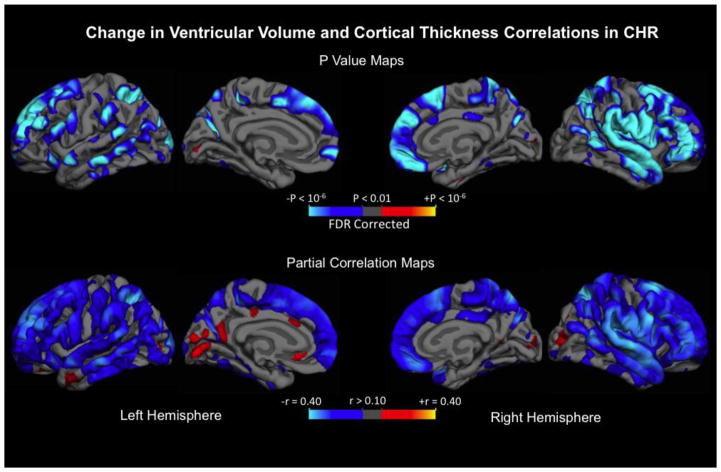
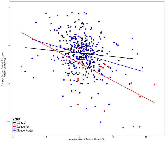
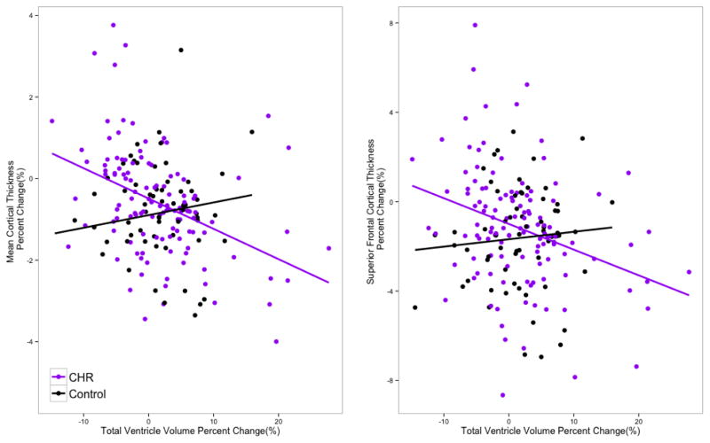
In a recent prospective longitudinal neuroimaging study, clinical high-risk (CHR) individuals who later developed full-blown psychosis showed an accelerated rate of gray matter thinning in superior and medial prefrontal cortex (PFC) and expansion of the ventricular system after applying a stringent correction for multiple comparisons. Although cortical and subcortical volume loss and enlarged ventricles are well characterized structural brain abnormalities among patients with schizophrenia, no prior study has evaluated whether these progressive changes of neuroanatomical indicators are linked in time prior to onset of psychosis. Therefore, we investigated the relationship between the changes in cortical gray matter thickness and ventricular volume using the longitudinal neuroimaging data from the North American Prodrome Longitudinal Study at the whole-brain level. The results showed that ventricular expansion is linked in time to progressive reduction of gray matter, rather than to structural changes in proximal subcortical regions, in a broadly distributed set of cortical regions among CHR youth, including superior, medial, lateral, and inferior PFC, superior temporal gyrus, and parietal cortices. In contrast, healthy controls did not show the same pattern of associations. The main findings were further replicated using a third assessment wave of MRI scans in a subset of study participants who were followed for an additional year. These findings suggest that the gray matter regions exhibiting aberrant rates of thinning in relation to psychosis risk are not limited to the PFC regions that survived the statistical threshold in our primary study, but also extend to other cortical regions previously implicated in schizophrenia.
Keywords: CHR; MRI; Prodromal; Psychosis; Schizophrenia; Ventricle.
Copyright © 2017 Elsevier B.V. All rights reserved.
Conflict of interest statement
The authors have declared that there are no conflicts of interest in relation to the subject of this study. Dr. Cannon reports that he is a consultant to the Los Angeles County Department of Mental Health and to Boerhinger Ingelheim Pharmaceutical and is a co-inventor (with the other NAPLS investigators) on a pending patent of a blood-based predictive biomarker for psychosis..
Figures
References
-
- Borgwardt SJ, McGuire PK, Aston J, Gschwandtner U, Pflüger MO, Stieglitz RD, Radue EW, Riecher-Rössler A. Reductions in frontal, temporal and parietal volume associated with the onset of psychosis. Schizophrenia Research. 2008;106:108–114. - PubMed
-
- Cannon TD, Chung Y, He G, Sun D, Jacobson A, van Erp TGM, McEwen S, Addington J, Bearden CE, Cadenhead K, Cornblatt B, Mathalon DH, McGlashan T, Perkins D, Jeffries C, Seidman LJ, Tsuang M, Walker E, Woods SW, Heinssen R North American Prodrome Longitudinal Study Consortium. Progressive reduction in cortical thickness as psychosis develops: a multisite longitudinal neuroimaging study of youth at elevated clinical risk. Biological Psychiatry. 2015;77:147–157. - PMC - PubMed
-
- Cannon TD, Sun F, McEwen SJ, Papademetris X, He G, van Erp TGM, Jacobson A, Bearden CE, Walker E, Hu X, Zhou L, Seidman LJ, Thermenos HW, Cornblatt B, Olvet DM, Perkins D, Belger A, Cadenhead K, Tsuang M, Mirzakhanian H, Addington J, Frayne R, Woods SW, McGlashan TH, Constable RT, Qiu M, Mathalon DH, Thompson P, Toga AW. Reliability of neuroanatomical measurements in a multisite longitudinal study of youth at risk for psychosis. Hum Brain Mapp. 2013;35:2424–2434. - PMC - PubMed
Publication types
MeSH terms
Grants and funding
- U01 MH082022/MH/NIMH NIH HHS/United States
- U01 MH081984/MH/NIMH NIH HHS/United States
- UL1 TR001863/TR/NCATS NIH HHS/United States
- U01 MH081902/MH/NIMH NIH HHS/United States
- P50 MH080272/MH/NIMH NIH HHS/United States
- U01 MH081988/MH/NIMH NIH HHS/United States
- P50 MH066286/MH/NIMH NIH HHS/United States
- U01 MH076989/MH/NIMH NIH HHS/United States
- K01 MH099431/MH/NIMH NIH HHS/United States
- U01 MH081928/MH/NIMH NIH HHS/United States
- U01 MH081857/MH/NIMH NIH HHS/United States
- U01 MH082004/MH/NIMH NIH HHS/United States
- U01 MH081944/MH/NIMH NIH HHS/United States
LinkOut - more resources
Full Text Sources
Other Literature Sources
Medical
Miscellaneous
