

Case Reports
doi: 10.1136/bcr-2016-219119.
Codfish vertebra sign
Affiliations
- PMID: 28246115
- PMCID: PMC5337632
- DOI: 10.1136/bcr-2016-219119
Item in Clipboard
Case Reports
Codfish vertebra sign
BMJ Case Rep.
.
No abstract available
Conflict of interest statement
Figures
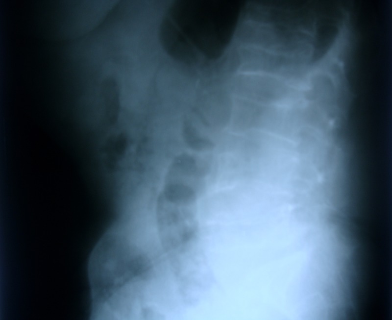
Lateral X-ray film of the lumbar spine, which reveals biconcavity of the lumbar vertebrae and osteopenia.
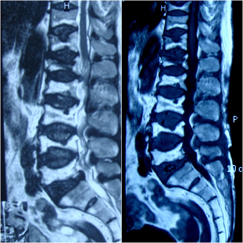
Sagittal T1-weighted (right) and T2-weighted (left) MRI of the lumbosacral spine. The D10–12 vertebrae are also seen. All vertebral bodies show biconcave deformity of their endplates with areas of rounded depressions and impressions, different signal intensities of the vertebral body and bone marrow, and loss of more than 40% of the vertebral height. This is severe osteoporosis showing different stages of bony collapse.
References
-
- Murphy WA Jr, DiVito DM. Fuller Albright, postmenopausal osteoporosis, and fish vertebrae. Radiology 2013;268:323–6. doi:10.1148/radiol.13122478 - DOI - PubMed
-
- Ntagiopoulos PG, Moutzouris DA, Manetas S. The ‘fish-vertebra’ sign. Emerg Med J 2007;24:674–5. doi:10.1136/emj.2006.039131 - DOI - PMC - PubMed
-
- Yamato M, Nishimura G, Kuramochi E et al. MR appearance at different ages of osteoporotic compression fractures of the vertebrae. Radiat Med 1998;16:329–34. - PubMed
Publication types
MeSH terms
LinkOut - more resources
Full Text Sources
Other Literature Sources