

Defining the enzymatic pathway for polymorphic O-glycosylation of the pneumococcal serine-rich repeat protein PsrP
- PMID: 28246170
- PMCID: PMC5391752
- DOI: 10.1074/jbc.M116.770446
Defining the enzymatic pathway for polymorphic O-glycosylation of the pneumococcal serine-rich repeat protein PsrP
Abstract
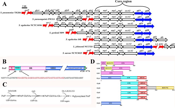
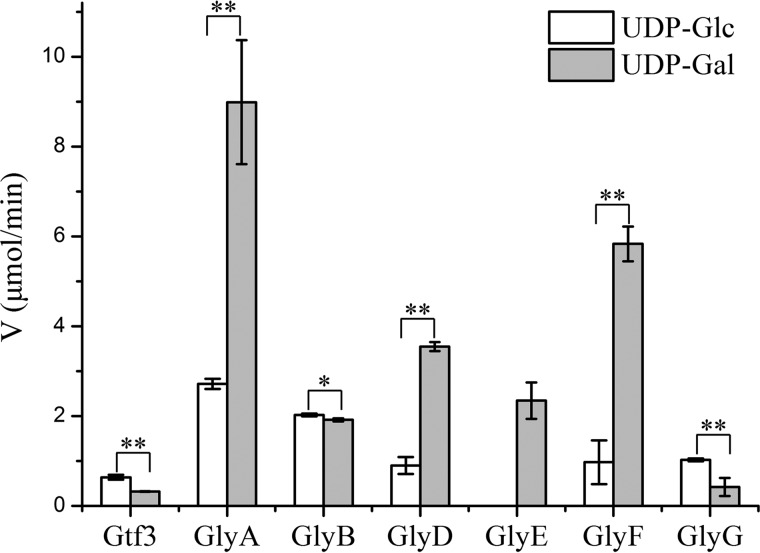
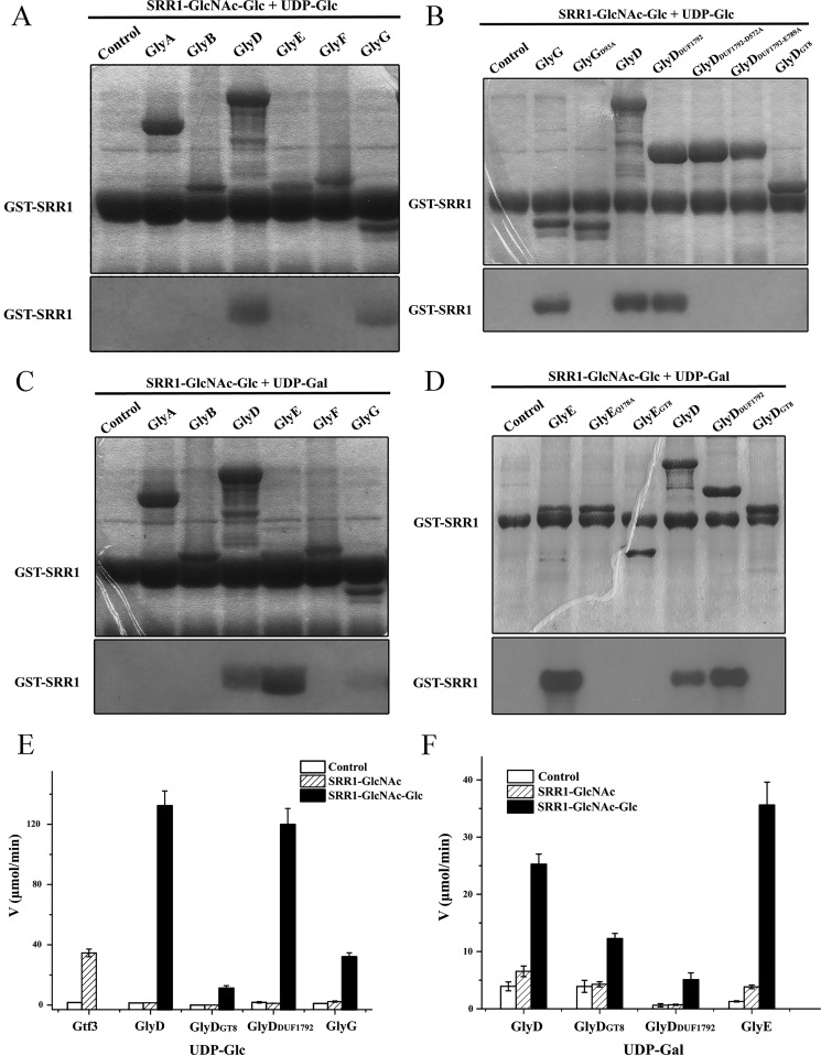
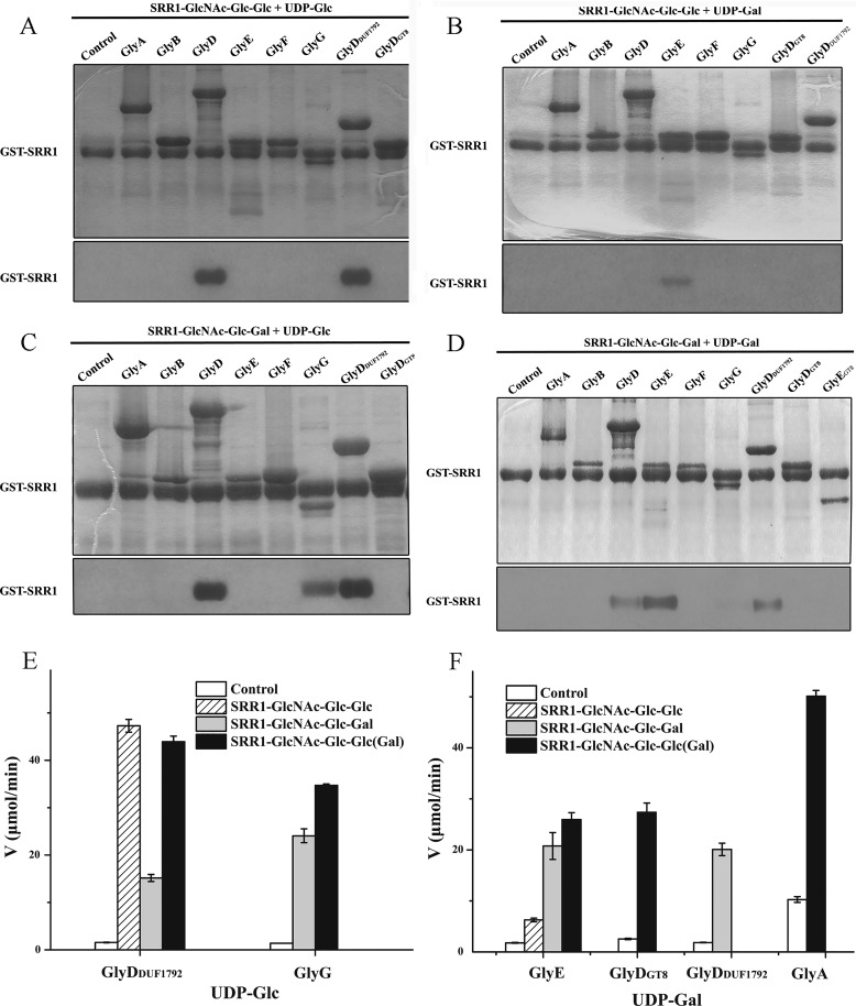
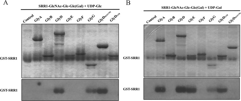
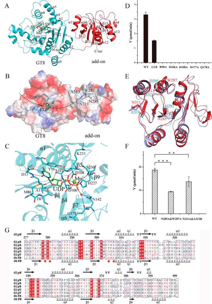
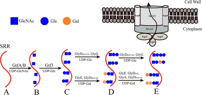
Protein O-glycosylation is an important post-translational modification in all organisms, but deciphering the specific functions of these glycans is difficult due to their structural complexity. Understanding the glycosylation of mucin-like proteins presents a particular challenge as they are modified numerous times with both the enzymes involved and the glycosylation patterns being poorly understood. Here we systematically explored the O-glycosylation pathway of a mucin-like serine-rich repeat protein PsrP from the human pathogen Streptococcus pneumoniae TIGR4. Previous works have assigned the function of 3 of the 10 glycosyltransferases thought to modify PsrP, GtfA/B, and Gtf3 as catalyzing the first two reactions to form a unified disaccharide core structure. We now use in vivo and in vitro glycosylation assays combined with hydrolytic activity assays to identify the glycosyltransferases capable of decorating this core structure in the third and fourth steps of glycosylation. Specifically, the full-length GlyE and GlyG proteins and the GlyD DUF1792 domain participate in both steps, whereas full-length GlyA and the GlyD GT8 domain catalyze only the fourth step. Incorporation of different sugars to the disaccharide core structure at multiple sites along the serine-rich repeats results in a highly polymorphic product. Furthermore, crystal structures of apo- and UDP-complexed GlyE combined with structural analyses reveal a novel Rossmann-fold "add-on" domain that we speculate to function as a universal module shared by GlyD, GlyE, and GlyA to forward the peptide acceptor from one enzyme to another. These findings define the complete glycosylation pathway of a bacterial glycoprotein and offer a testable hypothesis of how glycosyltransferase coordination facilitates glycan assembly.
Keywords: Streptococcus pneumoniae; bacterial pathogenesis; crystal structure; enzyme; glycosylation; glycosyltransferase; polymorphism; serine-rich repeat protein.
© 2017 by The American Society for Biochemistry and Molecular Biology, Inc.
Conflict of interest statement
The authors declare that they have no conflicts of interest with the contents of this article
Figures
Similar articles
-
Glycosyltransferases within the psrP Locus Facilitate Pneumococcal Virulence.J Bacteriol. 2021 Mar 8;203(7):e00389-20. doi: 10.1128/JB.00389-20. Print 2021 Mar 8. J Bacteriol. 2021. PMID: 33468592 Free PMC article.
-
Structure of a novel O-linked N-acetyl-D-glucosamine (O-GlcNAc) transferase, GtfA, reveals insights into the glycosylation of pneumococcal serine-rich repeat adhesins.J Biol Chem. 2014 Jul 25;289(30):20898-907. doi: 10.1074/jbc.M114.581934. J Biol Chem. 2014. PMID: 24936067 Free PMC article.
-
Engineering and Dissecting the Glycosylation Pathway of a Streptococcal Serine-rich Repeat Adhesin.J Biol Chem. 2016 Dec 30;291(53):27354-27363. doi: 10.1074/jbc.M116.752998. J Biol Chem. 2016. PMID: 28039332 Free PMC article.
-
Glycosyltransferase-mediated Sweet Modification in Oral Streptococci.J Dent Res. 2015 May;94(5):659-65. doi: 10.1177/0022034515574865. Epub 2015 Mar 9. J Dent Res. 2015. PMID: 25755271 Free PMC article. Review.
-
Sugar and Spice Make Bacteria Not Nice: Protein Glycosylation and Its Influence in Pathogenesis.J Mol Biol. 2016 Aug 14;428(16):3206-3220. doi: 10.1016/j.jmb.2016.04.013. Epub 2016 Apr 20. J Mol Biol. 2016. PMID: 27107636 Review.
Cited by
-
Identification of lthB, a Gene Encoding a Putative Glycosyltransferase Family 8 Protein Required for Leptothrix Sheath Formation.Appl Environ Microbiol. 2023 Apr 26;89(4):e0191922. doi: 10.1128/aem.01919-22. Epub 2023 Mar 23. Appl Environ Microbiol. 2023. PMID: 36951572 Free PMC article.
-
Membrane trafficking of the bacterial adhesin GspB and the accessory Sec transport machinery.J Biol Chem. 2019 Feb 1;294(5):1502-1515. doi: 10.1074/jbc.RA118.005657. Epub 2018 Dec 4. J Biol Chem. 2019. PMID: 30514759 Free PMC article.
-
Streptococcal Serine-Rich Repeat Proteins in Colonization and Disease.Front Microbiol. 2020 Oct 30;11:593356. doi: 10.3389/fmicb.2020.593356. eCollection 2020. Front Microbiol. 2020. PMID: 33193266 Free PMC article. Review.
-
Transcriptional organization of pneumococcal psrP-secY2A2 and impact of GtfA and GtfB deletion on PsrP-associated virulence properties.Microbes Infect. 2017 Jun;19(6):323-333. doi: 10.1016/j.micinf.2017.04.001. Epub 2017 Apr 10. Microbes Infect. 2017. PMID: 28408270 Free PMC article.
-
Serine-rich repeat protein adhesins from Lactobacillus reuteri display strain specific glycosylation profiles.Glycobiology. 2019 Jan 1;29(1):45-58. doi: 10.1093/glycob/cwy100. Glycobiology. 2019. PMID: 30371779 Free PMC article.
References
-
- Nothaft H., and Szymanski C. M. (2010) Protein glycosylation in bacteria: sweeter than ever. Nat. Rev. Microbiol. 8, 765–778 - PubMed
-
- Schwarz F., and Aebi M. (2011) Mechanisms and principles of N-linked protein glycosylation. Curr. Opin Struct. Biol. 21, 576–582 - PubMed
-
- Apweiler R., Hermjakob H., and Sharon N. (1999) On the frequency of protein glycosylation, as deduced from analysis of the SWISS-PROT database. Biochim. Biophys. Acta 1473, 4–8 - PubMed
-
- Ohtsubo K., and Marth J. D. (2006) Glycosylation in cellular mechanisms of health and disease. Cell 126, 855–867 - PubMed
Publication types
MeSH terms
Substances
Associated data
- Actions
- Actions
LinkOut - more resources
Full Text Sources
Other Literature Sources
Molecular Biology Databases
