Review
. 2017 May 1;9(5):a027714.
doi: 10.1101/cshperspect.a027714.

Membrane Currents, Gene Expression, and Circadian Clocks

Affiliations
Review

Membrane Currents, Gene Expression, and Circadian Clocks

Charles N Allen et al. Cold Spring Harb Perspect Biol. .

Abstract

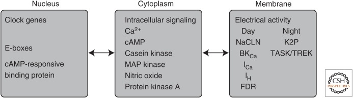
Neuronal circadian oscillators in the mammalian and Drosophila brain express a circadian clock comprised of interlocking gene transcription feedback loops. The genetic clock regulates the membrane electrical activity by poorly understood signaling pathways to generate a circadian pattern of action potential firing. During the day, Na+ channels contribute an excitatory drive for the spontaneous activity of circadian clock neurons. Multiple types of K+ channels regulate the action potential firing pattern and the nightly reduction in neuronal activity. The membrane electrical activity possibly signaling by changes in intracellular Ca2+ and cyclic adenosine monophosphate (cAMP) regulates the activity of the gene clock. A decline in the signaling pathways that link the gene clock and neural activity during aging and disease may weaken the circadian output and generate significant impacts on human health.


Figures

Figure 1.
Figure 1.
Cellular signaling domains mediating the generation of circadian rhythms in suprachiasmatic nucleus neurons. Molecular timekeeping signals generated in the nucleus are transmitted via intracellular signaling pathways to alter the activity of ion channels located in the neuronal membrane. Changes in the membrane electrical potential are communicated back to the molecular clock to synchronize and stabilize molecular rhythms. NACLN, Voltage-insensitive nonselective cation channel; cAMP, cyclic adenosine monophosphate; BKCa, large conductance calcium-activated potassium channel; ICa, voltage-gated calcium channels; IH, hyperpolarization-activated channel; K2P, two-pore domain potassium channel; TASK, TWIK (two-pore domain weak inward rectifying potassium channel)-related acid-sensitive K+ channel; TREK, TWIK-related potassium channel; FDR, fast delayed rectifier.
Figure 2.
Figure 2.
Intracellular calcium rhythms are present in mouse and Drosophila pacemaker neurons. (A) Long-duration intracellular calcium levels recorded cameleon expressed in a ventral SCN neuron (red dots). Simultaneous recording of multiunit activity was performed using a multielectrode array. The peak of the calcium rhythm of neuron preceded the peak of the multiunit activity by 6 h. Asterisks indicate the timing of the culture medium exchange, during which the firing frequency became temporarily unstable. (Panel A from Ikeda et al. 2003; reprinted, with permission, from Elsevier © 2003.) (B) Average calcium transients in five identified groups of circadian pacemaker neurons in the Drosophila brain. The calcium transients were measured using GCaMP6s. Note that the calcium levels peak at different times of the day in different neuron populations. (Panel B from Liang et al. 2016; reprinted, with permission, from AAAS © 2016.)
Figure 3.
Figure 3.
Suprachiasmatic nucleus (SCN) neuronal activity in a mouse model of Huntington’s disease. The spontaneous action potential firing frequency is reduced in the SCN of mice expressing the entire human huntingtin gene (HTT) with 97 mixed CAA-CAG repeats (BACHD). The current-clamp recording technique was used in the cell-attached configuration to record the spontaneous firing rate (SFR) of dorsal SCN neurons during the day (ZT4-6, top row) and night (ZT16-18, bottom row). The reduced excitability of SCN neurons in the BACHD mice are consistent with the hypothesis that reduced circadian output signals from the SCN contribute to the phenotypes observed in the BACHD mice.

References

    1. Akiyama M, Minami Y, Nakajima T, Moriya T, Shibata S. 2001. Calcium and pituitary adenylate cyclase-activating polypeptide induced expression of circadian clock gene mPer1 in the mouse cerebellar granule cell culture. J Neurochem 78: 499–508. - PubMed
    1. Albrecht U, Sun ZS, Eichele G, Lee CC. 1997. A differential response of two putative mammalian circadian regulators, mper1 and mper2, to light. Cell 91: 1055–1064. - PubMed
    1. Albus H, Bonnefont X, Chaves I, Yasui A, Doczy J, van der Horst GT, Meijer JH. 2002. Cryptochrome-deficient mice lack circadian electrical activity in the suprachiasmatic nuclei. Curr Biol 12: 1130–1133. - PubMed
    1. Alvado L, Allen CN. 2008. Tetraethylammonium (TEA) increases the inactivation time constant of the transient K+ current in suprachiasmatic nucleus neurons. Brain Res 1221: 24–29. - PMC - PubMed
    1. Asai M, Yoshinobu Y, Kaneko S, Mori A, Nikaido T, Moriya T, Akiyama M, Shibata S. 2001. Circadian profile of Per gene mRNA expression in the suprachiasmatic nucleus, paraventricular nucleus, and pineal body of aged rats. J Neurosci Res 66: 1133–1139. - PubMed