Review
. 2018 Jan 2;10(1):a022194.
doi: 10.1101/cshperspect.a022194.

TGF-β Family Signaling in Epithelial Differentiation and Epithelial-Mesenchymal Transition

Affiliations
Review

TGF-β Family Signaling in Epithelial Differentiation and Epithelial-Mesenchymal Transition

Kaoru Kahata et al. Cold Spring Harb Perspect Biol. .

Abstract

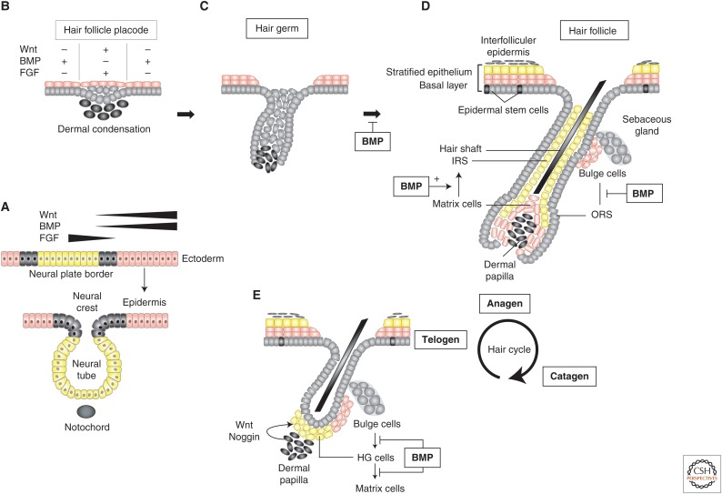
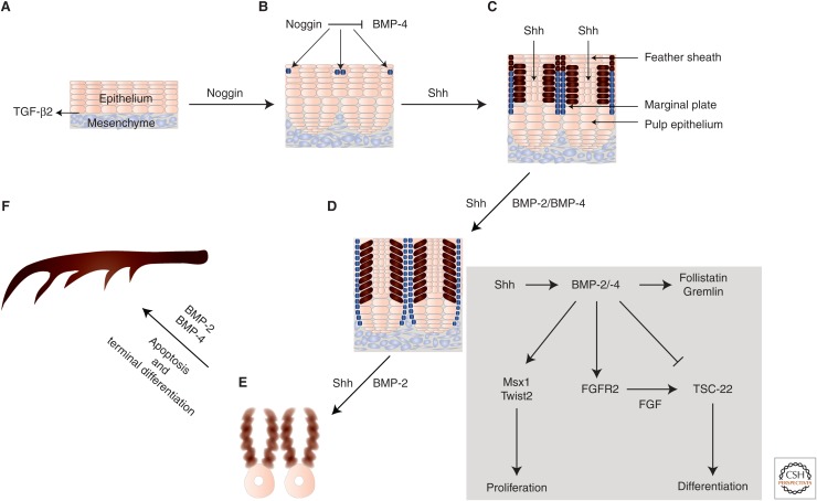
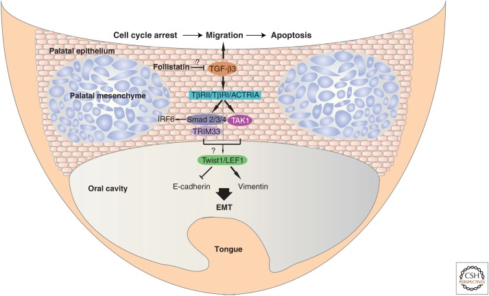
Epithelia exist in the animal body since the onset of embryonic development; they generate tissue barriers and specify organs and glands. Through epithelial-mesenchymal transitions (EMTs), epithelia generate mesenchymal cells that form new tissues and promote healing or disease manifestation when epithelial homeostasis is challenged physiologically or pathologically. Transforming growth factor-βs (TGF-βs), activins, bone morphogenetic proteins (BMPs), and growth and differentiation factors (GDFs) have been implicated in the regulation of epithelial differentiation. These TGF-β family ligands are expressed and secreted at sites where the epithelium interacts with the mesenchyme and provide paracrine queues from the mesenchyme to the neighboring epithelium, helping the specification of differentiated epithelial cell types within an organ. TGF-β ligands signal via Smads and cooperating kinase pathways and control the expression or activities of key transcription factors that promote either epithelial differentiation or mesenchymal transitions. In this review, we discuss evidence that illustrates how TGF-β family ligands contribute to epithelial differentiation and induce mesenchymal transitions, by focusing on the embryonic ectoderm and tissues that form the external mammalian body lining.


Figures

Figure 1.
Figure 1.
Roles of bone morphogenetic protein (BMP) signaling in epidermal development. (A) Ectoderm definition. Ectodermal cells (pink) are separated from the neural plate cells (yellow) by small patches of border cells (gray). These embryonic cell types are defined anatomically and based on the pathways that control their developmental fate. In ectodermal cells, Wnt-induced β-catenin signals are active, and BMP signaling is preserved and induces epidermis, whereas fibroblast growth factor (FGF) signaling promotes neural commitment in which Wnt–β-catenin signaling is silenced. Intermediate intensity of Wnt and BMP signals in the border region defines the limits of these embryonic territories. (BE) Hair follicle development and hair cycling. (B) Wnt–β-catenin (+) and FGF (+) signaling induce placode formation accompanied by dermal condensation underneath. BMP (+) signaling on either side of the hair follicle placode inhibits its formation. (C) Hair follicle placode “downgrowth” away from the surface of the skin and engulfment of dermal condensations at the tip of the hair germ that grows downward. (D) Proliferation and differentiation of bulge cells toward outer root sheath (ORS) cells are blocked by BMP signals, whereas BMP signals are required for terminal differentiation of matrix cells to inner root sheath (IRS) cells that generate the hair shaft. Hair follicle stem cells (HFSCs) are accommodated in the bulge and hair germ. Bulge cells remain in a quiescent stage of their cell cycle and BMP signaling contributes to their lengthy life span. Hair germ cells are more prone to be activated by dermal signals and contribute postnatally to hair follicle regeneration. The three phases of the postnatal hair cycle are also shown. (E) At the end of the telogen phase, the dermal papilla secretes noggin, which inhibits BMP signaling; BMPs inhibit bulge cell differentiation to hair germ (HG) cells and matrix cells as explained in D. Noggin-mediated inhibition of BMP signaling leads to activation of HG cells and induction of their differentiation into matrix cells, whereas Wnt promotes HG cell proliferation.
Figure 2.
Figure 2.
Branching and evolution of feathers. (A,B) TGF-β2 localizes in the border where epithelium and mesenchyme interact. The multilayered epidermis can be divided into epidermal ridges (dark blue cells) by the ridge-forming activator noggin and the ridge-forming inhibitor BMP-4. Noggin and BMP-4 are distributed in the mesenchyme (not shown) and their balanced action is crucial for normal specification of these primordial ridge cells. (B,C) High BMP-4 levels at the proliferative zone lead to the formation of a cylindrical structure by the epithelial cells. Within the epidermis, when the level of noggin is high, the epithelia gradually start to form multiple barb ridges (blue cells). The role and localization of the upstream Sonic Hedgehog (Shh) is also indicated. (D) Expression of BMP-2 and BMP-4 is regulated by Shh signaling. Bone morphogenetic proteins (BMPs) mediate a lateral inhibitory mechanism that leads to arrangement of feather buds in a periodic pattern. BMP signaling in the feather bud cells (brown) regulates the expression of FGFR2. Moreover, BMP induces the expression of transcriptional regulators Msx1 and Twist2 that drive feather bud proliferation. Follistatin and gremlin, secreted by feather interbud dermal cells moderate the autocrine action of BMP signaling via a feedback mechanism. TSC-22 expression is repressed by BMP signaling and induced by FGF signaling, and is required for feather cell differentiation. Marginal plate cells that express Shh and BMP-2 undergo apoptosis, and Shh-negative barb ridge cells continue proliferating. (E,F) Barb ridge cells express BMP-2 and BMP-4 and form the more differentiated structures known as barbules. Barbule plate cells form cilia and hooklets. Noggin activity toward the end of feather formation is reduced and leads to formation of the calamus without branches at the proximal end of the feather shaft. The site with higher BMP activity forms the bilateral symmetric feather. (The image has been inspired by the article of Yu et al. 2002.)
Figure 3.
Figure 3.
Molecular control of palatal development. TGF-β3 plays a crucial role in palatal development and closure. TGF-β3 signals through TβRII and TβRI receptors to stimulate the formation of Smad2–Smad3–Smad4 complexes. The ubiquitin ligase TRIM33 also participates in complexes with Smad2 and/or Smad3; cooperative actions between Smad, TRIM33, and TAK1 signaling may activate expression of the epithelial–mesenchymal transition (EMT) transcription factors Twist1 and LEF1 (note the “question mark,” as the latter mechanism is not firmly proven), which directly activate the expression of mesenchymal genes, such as the gene encoding vimentin, and suppress the expression of epithelial genes including the Cdh1 gene encoding E-cadherin during the palatal EMT. Smad complexes may also interact with IRF6 to regulate additional target genes during EMT. The possible involvement of follistatin, which may (note the “question mark”) indirectly limit TGF-β3 activity, and ActRI/ALK-2 as an alternative type I receptor, are also indicated to reflect the discussion in the main text. The sequence of events induced by TGF-β is epithelial cell cycle arrest, followed by cell migration and eventual apoptosis at the seam (top of the figure). (The image has been inspired by Bush and Jiang 2012.)

References

    1. Acloque H, Adams MS, Fishwick K, Bronner-Fraser M, Nieto MA. 2009. Epithelial–mesenchymal transitions: The importance of changing cell state in development and disease. J Clin Invest 119: 1438–1449. - PMC - PubMed
    1. Ahmed S, Liu CC, Nawshad A. 2007. Mechanisms of palatal epithelial seam disintegration by transforming growth factor (TGF) β3. Dev Biol 309: 193–207. - PMC - PubMed
    1. Amendt C, Schirmacher P, Weber H, Blessing M. 1998. Expression of a dominant negative type II TGF-β receptor in mouse skin results in an increase in carcinoma incidence and an acceleration of carcinoma development. Oncogene 17: 25–34. - PubMed
    1. Andersson O, Bertolino P, Ibanez CF. 2007. Distinct and cooperative roles of mammalian Vg1 homologs GDF1 and GDF3 during early embryonic development. Dev Biol 311: 500–511. - PubMed
    1. Andl T, Reddy ST, Gaddapara T, Millar SE. 2002. WNT signals are required for the initiation of hair follicle development. Dev Cell 2: 643–653. - PubMed

Publication types

Substances

LinkOut - more resources