Review
. 2017 Nov 1;9(11):a022269.
doi: 10.1101/cshperspect.a022269.

TGF-β Family Signaling in Connective Tissue and Skeletal Diseases

Affiliations
Review

TGF-β Family Signaling in Connective Tissue and Skeletal Diseases

Elena Gallo MacFarlane et al. Cold Spring Harb Perspect Biol. .

Abstract

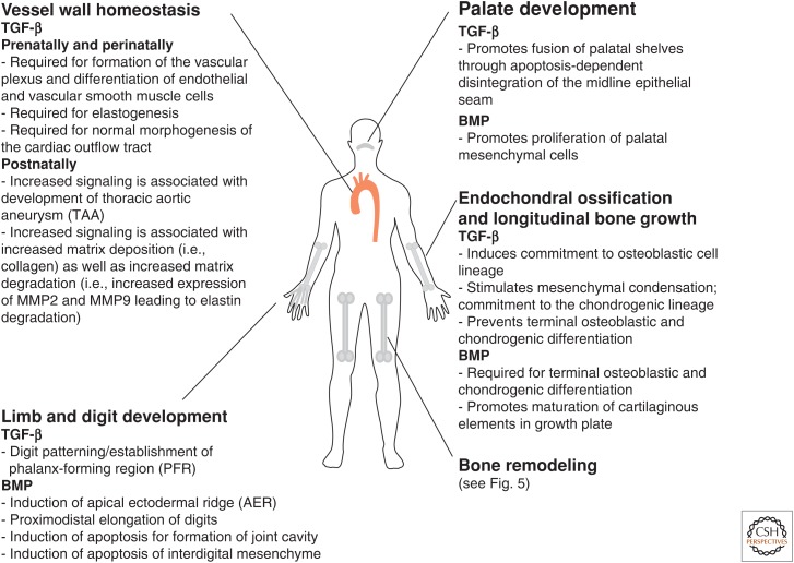
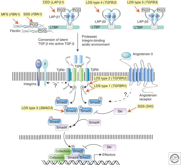
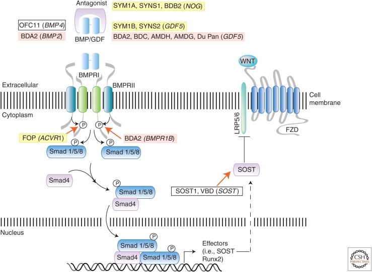
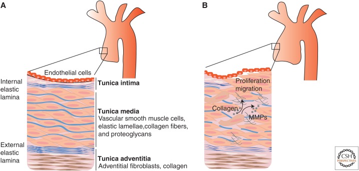
The transforming growth factor β (TGF-β) family of signaling molecules, which includes TGF-βs, activins, inhibins, and numerous bone morphogenetic proteins (BMPs) and growth and differentiation factors (GDFs), has important functions in all cells and tissues, including soft connective tissues and the skeleton. Specific TGF-β family members play different roles in these tissues, and their activities are often balanced with those of other TGF-β family members and by interactions with other signaling pathways. Perturbations in TGF-β family pathways are associated with numerous human diseases with prominent involvement of the skeletal and cardiovascular systems. This review focuses on the role of this family of signaling molecules in the pathologies of connective tissues that manifest in rare genetic syndromes (e.g., syndromic presentations of thoracic aortic aneurysm), as well as in more common disorders (e.g., osteoarthritis and osteoporosis). Many of these diseases are caused by or result in pathological alterations of the complex relationship between the TGF-β family of signaling mediators and the extracellular matrix in connective tissues.


Figures

Figure 1.
Figure 1.
Summary of the roles of transforming growth factor β (TGF-β) and bone morphogenetic protein (BMP) signaling in the development and homeostasis of connective tissue and the skeletal system.
Figure 2.
Figure 2.
Summary of the transforming growth factor β (TGF-β) signaling pathway and mutations causing connective tissue disorders. TGF-β molecules (TGF-β1, -β2, and -β3) are secreted in latent forms that are unable to interact with the receptors. In the latent form, dimeric TGF-β molecules are noncovalently associated with dimeric latency-associated peptides (LAPs), which are made from the same gene as the corresponding TGF-β molecule. This complex, which is referred to as the small latent complex (SLC), is then covalently linked by disulfide bonds to a member of the latent TGF-β binding protein (LTBP) family, to form the larger complex called large latent complex (LLC). The LLC complex interacts noncovalently with various components of the extrcellular matrix (ECM), including fibrillin microfibrils and integrins, the major adhesion molecule linking cells to the ECM. Both fibrillin and TGF-β (TGF-β1 and -β3, but not TGF-β2) contain an arginine-glycine-aspartic acid (RGD) domain that can bind integrin molecules. Cross talk between LTBP, fibrillin, and integrins is thought to be critical for proper localization, sequestration, and conversion of latent TGF-β to active TGF-β. Once activated, dimeric TGF-β binds to a tetrameric receptor complex formed by two type I (TβRI) and type II (TβRII) receptor subunits, leading to direct phosphorylation of R-Smads (Smad2 or Smad3), complex formation with co-Smad (Smad4), and induction or repression of target gene expression. Heterozygous inactivating mutations in both “positive” and “negative” regulators of this pathway have been identified as the cause of genetic disorders that are characterized by pathological alterations in the connective tissue due to misregulated expression of TGF-β gene targets (i.e., tissue metalloproteinases, collagen, integrins, and several other ECM components). The syndromes associated with these mutations, and the human gene mutated in each condition, are highlighted in yellow.
Figure 3.
Figure 3.
Summary of the bone morphogenetic protein (BMP) and growth and differentiation factor (GDF) signaling pathway and mutations causing connective tissue and skeletal disorders. The BMP signaling pathway is activated by extracellular ligands, such as BMPs and GDFs, through binding to type I (BMPRI) and type II (BMPRII) receptor complexes. Extracellular antagonists, such as noggin, can block ligand–receptor binding and pathway activation. Smad (shown) and non-Smad (not shown) mechanisms mediate BMP signal transduction to regulate transcriptional target genes, including sclerostin (SOST), a Wnt pathway inhibitor. Key components of the BMP pathway are shown to illustrate positions at which gene mutations act to cause specific human connective tissue and skeletal syndromes. Cross talk with the Wnt pathway is also shown. Specific disorders and their gene mutations (in parentheses), as discussed in text and Table 1, are shaded in yellow if the mutation enhances pathway signaling or in red if the pathway has diminished activation. White boxes indicate disorders for which the BMP pathway is either not directly affected or the functional consequence of the mutation is unknown. BDA2, Brachydactyly type A2; BDC, brachydactyly type C; AMDH, acromesomelic dysplasia Hunter–Thompson type; AMDG, acromesomelic dysplasia Grebe type; FOP, fibrodysplasia ossificans progressiva; VBD, Van Buchem disease.
Figure 4.
Figure 4.
Schematic representation of the vessel wall of major arteries. (A) The vessel wall of arteries is organized in layers, which are referred to as tunica intima, media, and adventitia. Endothelial cells line the lumen and are attached to a basement membrane. The connective tissue immediately below the endothelial cell layer is referred to as tunica intima. The internal elastic lamina separates the tunica intima from the tunica media, which contains vascular smooth muscle cells (VSMCs) and elastic fibers, organized in fenestrated sheets (also called lamellae). The external elastic lamina separates the tunica media from the tunica adventitia, which contains mostly fibroblasts and collagen, but no elastic fibers. (B) Arterial wall remodeling associated with aneurysm development include increased VSMC migration and proliferation (and associated loss of contractile function), increased secretion of matrix-degrading enzymes (such as matrix metalloproteinases [MMPs]), and increased deposition of collagen and fragmentation of elastic fibers. The ultimate effect of these processes is the replacement of the ordered and layered architecture of elastic lamellae and vascular smooth muscle cells with disorganized and weak connective tissue.
Figure 5.
Figure 5.
Roles of transforming growth factor β (TGF-β) and bone morphogenetic protein (BMP) signaling in bone homeostasis. Bone homeostasis is maintained through continuous cycles of bone resorption by osteoclasts and new bone formation by osteoblasts and osteocytes. Both TGF-β and BMP signaling participate in regulating this process. TGF-β can act as a chemoattractant and recruit both preosteoblast and preosteoclasts to areas of bone remodeling and also promotes proliferation, differentiation, and survival of osteoclasts. At low doses, TGF-β increases secretion of RANKL (receptor activator of NF-κB ligand) and suppresses expression of the inhibitor OPG (osteoprotegerin), thus promoting osteoclastogenesis by activating RANK signaling in preosteoclasts. At high doses, it suppresses RANKL and increases OPG expression and thus inhibits osteoclastogenesis. This biphasic effect limits excessive bone degradation in the presence of high levels of active TGF-β derived from conversion of latent TGF-β by proteases and acid secreted by osteoclasts during bone degradation. BMP signaling suppresses osteoclast differentiation by increasing expression of OPG in osteoblasts. It can, however, also promote bone resorption by increasing the expression of sclerostin (SOST), an inhibitor of Wnt signaling which is a pathway that normally promotes osteoblastogenesis and bone formation. M-CSF, Macrophage-colony stimulating factor.

References

    1. Ahn K, Mishina Y, Hanks MC, Behringer RR, Crenshaw EB III. 2001. BMPR-IA signaling is required for the formation of the apical ectodermal ridge and dorsal–ventral patterning of the limb. Development 128: 4449–4461. - PubMed
    1. Akiyama H. 2008. Control of chondrogenesis by the transcription factor Sox9. Mod Rheumatol 18: 213–219. - PubMed
    1. Akiyama H, Chaboissier MC, Martin JF, Schedl A, de Crombrugghe B. 2002. The transcription factor Sox9 has essential roles in successive steps of the chondrocyte differentiation pathway and is required for expression of Sox5 and Sox6. Genes Dev 16: 2813–2828. - PMC - PubMed
    1. Akiyama T, Marques G, Wharton KA. 2012. A large bioactive BMP ligand with distinct signaling properties is produced by alternative proconvertase processing. Sci Signal 5: ra28. - PMC - PubMed
    1. Akiyoshi S, Inoue H, Hanai J, Kusanagi K, Nemoto N, Miyazono K, Kawabata M. 1999. c-Ski acts as a transcriptional co-repressor in transforming growth factor-β signaling through interaction with Smads. J Biol Chem 274: 35269–35277. - PubMed

LinkOut - more resources