. 2017 Mar 1;144(5):830-836.
doi: 10.1242/dev.144063.

The assembly of developing motor neurons depends on an interplay between spontaneous activity, type II cadherins and gap junctions

Affiliations

The assembly of developing motor neurons depends on an interplay between spontaneous activity, type II cadherins and gap junctions

Karli Montague et al. Development. .

Abstract

A core structural and functional motif of the vertebrate central nervous system is discrete clusters of neurons or 'nuclei'. Yet the developmental mechanisms underlying this fundamental mode of organisation are largely unknown. We have previously shown that the assembly of motor neurons into nuclei depends on cadherin-mediated adhesion. Here, we demonstrate that the emergence of mature topography among motor nuclei involves a novel interplay between spontaneous activity, cadherin expression and gap junction communication. We report that nuclei display spontaneous calcium transients, and that changes in the activity patterns coincide with the course of nucleogenesis. We also find that these activity patterns are disrupted by manipulating cadherin or gap junction expression. Furthermore, inhibition of activity disrupts nucleogenesis, suggesting that activity feeds back to maintain integrity among motor neurons within a nucleus. Our study suggests that a network of interactions between cadherins, gap junctions and spontaneous activity governs neuron assembly, presaging circuit formation.

Keywords: Brainstem; Cadherins; Cell adhesion; Chick; Gap junctions; Motor neurons; Spontaneous activity.


Conflict of interest statement

Competing interests

The authors declare no competing or financial interests.

Figures
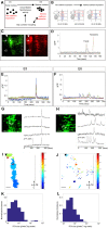
Fig. 1.
Fig. 1.
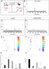
Spontaneous activity of facial motor neurons shows different patterns at E5 and E6. (A) Schematic model of the role of nucleogenesis. (B) Schematic of rhombomeres (r) 4-6 of the chick hindbrain in ‘open book’ prep; the floor plate is positioned medially (grey bar), the facial nerve and otic vesicle (grey circle) are on the left. Facial and abducens motor neurons are indicated by red and blue circles, respectively. (C) z-stack confocal images of hindbrain with GCaMP-electroporated facial motor neurons (left) or retrogradely labelled with fluorescent rhodamine-dextrans (right). Dashed line indicates the floor plate; arrowhead indicates lateral edge of the neuroepithelium. The mature coalescing nucleus is shown by the asterisk. (D) E5 example time series showing normalised ΔF/F GCaMP fluorescence for individual facial and abducens neurons (n=3 preparations). (E,F) E5 (E) and E6 (F) example time series for ΔF/F of facial motor neurons [n=16 preparations (E) and n=17 preparations (F)]. (G,H) E5 (G) and E6 (H) examples of identified GCaMP-expressing facial motor neurons (left) with individual ΔF/F traces (right). Circles indicate the positions of cells from which traces were recorded. (I,J) E5 (I) and E6 (J) phase maps derived from individual time series showing active ROIs within the field of view of the facial motor nucleus. Colour represents time of activity peak (in seconds), with colder colours representing earlier events in the time series and warmer colours representing later events. Dashed line indicates floor plate; arrowhead indicates lateral edge of neuroepithelium. L-M, lateral-medial axis; R-C, rostral-caudal axis. (K,L) E5 (K) and E6 (L) plots of ROI size. The x-axis is the size of the region of interest (ROI in pixels2; 100 pixels2=25 μm2) on a log scale; y-axis is the normalised incidence for each active ROI size, binned. Scale bars: 50 µm (C); 10 µm (G,H); 40 µm (I,J).
Fig. 2.
Fig. 2.
Perturbing cadherin-dependent interactions disrupts motor neuron spontaneous activity. (A) Schematic model of perturbation of cadherin-dependent interactions. (B-D) E5 example time series for ΔF/F of facial motor neurons (GCaMP control, NΔ390/GCaMP, Cad20 siRNA/GCaMP as labelled). Datasets are n=11 for NΔ390 and n=4 for Cad20 siRNA. (E,F) NΔ390 (E) and Cad20 (F) siRNA-electroporated hindbrain example phase maps, as in Fig. 1. Scale bars: 40 µm. (G) Frequency histogram showing mean number of bursts per minute for individual neurons. Compared with E5 controls, the frequency for E6 controls and NΔ390 is significantly higher (**P<0.01 and *P<0.05, respectively). Compared with E6 controls, the frequency for NΔ390, Cad20 siRNA and Cx43 siRNA was significantly lower (*P<0.05, **P<0.01 and *P<0.05, respectively). (H) Histogram showing ΔF/F amplitude under conditions as labelled. Amplitude is significantly lower for E6 and for Cx43 siRNA than for E5 (*P<0.05). Datasets for G and H are: E5 n=256 cells, E6 n=271 cells, NΔ390 n=150 cells, Cad20 siRNA n=43 cells, Cx43 siRNA n=56 cells.
Fig. 3.
Fig. 3.
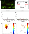
Perturbing gap junction-dependent interactions disrupts motor neuron spontaneous activity. (A) Single confocal section showing retrogradely labelled facial motor neurons (green) and whole-mount immunofluorescence for the gap junction protein Cx43 (red). (B) Schematic model of perturbation of gap junction-dependent interactions. (C,D) E5 example time series for ΔF/F of facial motor neurons for GCaMP control (C) and Cx43 siRNA/GCaMP (D). n=4 preparations. (E,F) E5 control (E) and Cx43 siRNA-electroporated (F) hindbrain example phase maps, labelled as in Fig. 1. Scale bars: 7 µm (A); 35 µm (E,F).
Fig. 4.
Fig. 4.
The effect of chronic activity inhibition on nucleogenesis and cadherin expression. (A) Schematic model of perturbation of spontaneous activity. (B) Schematic of an E6 transverse hindbrain section showing the mediolateral (L-M) location of facial (VII) and abducens (VI) motor neurons, and pattern of Cad13/Cad20 expression. (C,D) E6 transverse hindbrain sections immunostained for the motor neuron-specific marker Islet 1 (dark brown), showing the facial (single asterisk) and the abducens (double asterisk) motor nuclei. In situ hybridisation for either Cad20 (C) or Cad13 (D) was carried out on the same sections. Left: control, vehicle-treated embryos (Tyrode's solution); right: calcium channel blocker-treated embryos. Scale bars: 10 µm. (E) Quantification of facial motor nucleus coalescence in embryos as labelled. The proportion of facial motor neurons with no juxtaposing motor neurons increases significantly (P<0.01) following activity inhibition (n=3 experiments, 8-10 embryos per experiment).

References

    1. Astick M., Tubby K., Mubarak W. M., Guthrie S. and Price S. R. (2014). Central topography of cranial motor nuclei controlled by differential cadherin expression. Curr. Biol. 24, 2541-2547. 10.1016/j.cub.2014.08.067 - DOI - PMC - PubMed
    1. Benjumeda I., Escalante A., Law C., Morales D., Chauvin G., Muca G., Coca Y., Marquez J., Lopez-Bendito G., Kania A. et al. (2013). Uncoupling of EphA/ephrinA signaling and spontaneous activity in neural circuit wiring. J. Neurosci. 33, 18208-18218. 10.1523/JNEUROSCI.1931-13.2013 - DOI - PMC - PubMed
    1. Chen T.-W., Wardill T. J., Sun Y., Pulver S. R., Renninger S. L., Baohan A., Schreiter E. R., Kerr R. A., Orger M. B., Jayaraman V. et al. (2013). Ultrasensitive fluorescent proteins for imaging neuronal activity. Nature 499, 295-300. 10.1038/nature12354 - DOI - PMC - PubMed
    1. Demireva E. Y., Shapiro L. S., Jessell T. M. and Zampieri N. (2011). Motor neuron position and topographic order imposed by beta- and gamma-catenin activities. Cell 147, 641-652. 10.1016/j.cell.2011.09.037 - DOI - PMC - PubMed
    1. Feller M. B., Wellis D. P., Stellwagen D., Werblin F. S. and Shatz C. J. (1996). Requirement for cholinergic synaptic transmission in the propagation of spontaneous retinal waves. Science 272, 1182-1187. 10.1126/science.272.5265.1182 - DOI - PubMed

Publication types