. 2017 Mar 14;114(11):E2253-E2262.
doi: 10.1073/pnas.1616892114. Epub 2017 Feb 28.

Synaptic vesicle glycoprotein 2C (SV2C) modulates dopamine release and is disrupted in Parkinson disease

Affiliations

Synaptic vesicle glycoprotein 2C (SV2C) modulates dopamine release and is disrupted in Parkinson disease

Amy R Dunn et al. Proc Natl Acad Sci U S A. .

Abstract

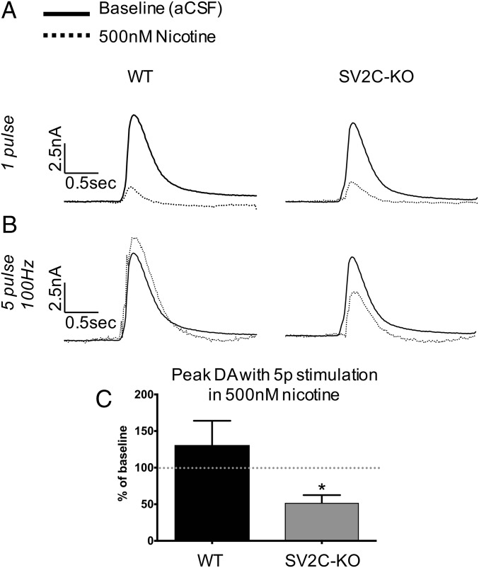
Members of the synaptic vesicle glycoprotein 2 (SV2) family of proteins are involved in synaptic function throughout the brain. The ubiquitously expressed SV2A has been widely implicated in epilepsy, although SV2C with its restricted basal ganglia distribution is poorly characterized. SV2C is emerging as a potentially relevant protein in Parkinson disease (PD), because it is a genetic modifier of sensitivity to l-DOPA and of nicotine neuroprotection in PD. Here we identify SV2C as a mediator of dopamine homeostasis and report that disrupted expression of SV2C within the basal ganglia is a pathological feature of PD. Genetic deletion of SV2C leads to reduced dopamine release in the dorsal striatum as measured by fast-scan cyclic voltammetry, reduced striatal dopamine content, disrupted α-synuclein expression, deficits in motor function, and alterations in neurochemical effects of nicotine. Furthermore, SV2C expression is dramatically altered in postmortem brain tissue from PD cases but not in Alzheimer disease, progressive supranuclear palsy, or multiple system atrophy. This disruption was paralleled in mice overexpressing mutated α-synuclein. These data establish SV2C as a mediator of dopamine neuron function and suggest that SV2C disruption is a unique feature of PD that likely contributes to dopaminergic dysfunction.

Keywords: Parkinson disease; SV2C; dopamine; synaptic vesicles; α-synuclein.


Conflict of interest statement

The authors declare no conflict of interest.

Figures

Fig. 1.
Fig. 1.
Polyclonal SV2C antibody characterization. We generated polyclonal SV2C antibodies. (A) SV2C antibody recognizes SV2C but does not recognize SV2A or SV2B in transfected HEK293 cells. Preincubating the SV2C antibody with the immunizing peptide (antigen blocking) eliminates antibody reactivity. (B) SV2C is highly colocalized with the dopaminergic marker TH in the mouse SNpc and dSTR. (Scale bars, 20 μm.) (C) Knocking down endogenous SV2C in N2a cells (which have no endogenous SV2A or SV2B) using shRNA reduces mSV2C antibody reactivity. (D) Sagittal DAB-stained sections showing ubiquitous SV2A and SV2B expression compared with SV2C expression enriched in the basal ganglia using the mSV2C antibody. Black arrowheads indicate areas of highest SV2C staining in the mouse brain: midbrain, dSTR, and ventral pallidum. Each sagittal image was compiled from a series of eight micrographs taken across a single section of tissue.
Fig. 2.
Fig. 2.
SV2C expression in mouse models of PD. (A) Striatal TH is 68% reduced after a 4× 15 mg/kg dose of MPTP (n = 6, P < 0.01). Striatal SV2C is reduced after MPTP (P < 0.05). Representative immunohistochemical staining after MPTP reveals that striatal SV2C expression patterns are unchanged. (B) Striatal SV2C levels are slightly, but not significantly, reduced in aged VMAT2-LO animals. SV2C expression patterns remain unaltered, as shown by representative immunohistochemistry in the dSTR. (C) An increase of SV2C-positive punctate staining is observed in the striatum of A53T-OE mice as compared with WT mice, particularly in the periventricular mediodorsal striatum. Clusters of punctate SV2C are found distributed sparsely in the striatum of A53T-OE mice. (Insets) Micrographs were taken at 2.5× and 40× magnification.
Fig. 3.
Fig. 3.
Neurochemical characterization of SV2C-KO mice. (A) PCR genotyping of SV2C-KO mice generated using the EUCOMM knockout-first allele. This construct allowed the generation of several useful lines of mice: a preliminary knockout animal with a cassette creating an frt-flanked gene trap inserted into the second exon (KOF), a line with a floxed exon following a cross of KOF animals with a Flp-recombinase+ line, and finally SV2C-KO mice following a cross with a nestin-driven Cre-recombinase+ line. Animals used for experiments are denoted by a superscript 1 (WT control) and 2 (SV2C-KO). (B) SV2C knockout does not result in altered expression of SV2A, SV2B, TH, DAT, or synaptophysin in the striatum. (C) Genetic deletion of SV2C ablates mSV2C antibody reactivity in the dSTR and midbrain. The dotted line delineates the dorsolateral striatum where electrochemical recordings were taken (Figs. 6 and 7). (D and E) SV2C knockout results in a 33% reduction of dopamine content (D) and a 19% reduction in the dopamine metabolite DOPAC (E) in the dSTR (n = 7, **P < 0. 01). (F) SV2C knockout does not result in a reduction in dopaminergic synaptic density in the dSTR. ns, not significant. (Scale bars, 500 µm in C and 20 μm in F.)
Fig. 4.
Fig. 4.
An association between SV2C and α-synuclein. (A) Midbrain homogenates from SV2C-KO animals have a 31% decrease in monomeric (15-kD) α-synuclein (n = 4–6, P = 0.06) and a 30-fold increase in multimeric (90-kD) α-synuclein (n = 4–6, P < 0.01). (B) α-Synuclein coimmunoprecipitates with SV2C in WT mouse striatal homogenates. TH, a cytosolic protein, and DAT, a membrane-associated protein, were included as negative controls. VMAT2 was included as a vesicular protein control. Synaptotagmin-1 (STG-1), which binds to SV2C, was used as a positive control. (C) Further confirmation of α-synuclein and SV2C coimmunoprecipitation. A non–antibody-bound immunoprecipitation (No AB) column served as negative control. Striatal homogenates from multiple animals were pooled and aliquoted to form the input for all three immunoprecipitations. (D and E) By immunohistochemistry, the patterns of the expression of SV2C-positive puncta are not similar to those of SV2A (D) or α-synuclein (E).
Fig. 5.
Fig. 5.
Altered motor behavior of SV2C-KO mice. (A) SV2C-KO animals have an ∼10% reduction in stride length as measured by a gait analysis assay (*P < 0.05). (B) Genetic deletion of SV2C does not result in impairment on the rotorod test. (C) SV2C-KO animals display a 23% reduction in total ambulations in 24-h locomotor activity monitoring (n = 7 or 8; *P < 0.05). (D) The reduction in locomotor activity apparently is driven by an ablation of a circadian peak in activity at the end of the active period. The shaded region in D indicates the dark phase.
Fig. 6.
Fig. 6.
Electrochemical measurement of stimulated dopamine release in SV2C-KO mice. Using FSCV, we measured dopamine release stimulated by a single electrical pulse in the dSTR. Genetically ablating SV2C reduces dopamine release by 32% as shown by a representative color blot (A), as quantified over all recording sites in each animal (n = 9; *P < 0.05) (B), and as represented by respective current traces (C).
Fig. 7.
Fig. 7.
Altered neurochemical effect of nicotine after genetic ablation of SV2C as measured by FSCV. (A) Current traces showing dopamine release at baseline and in the presence of 500 nM nicotine. Nicotine normally reduces dopamine release elicited by a one-pulse stimulation to about 10% of baseline in both WT and SV2C-KO striatum. (B) Five-pulse stimulations in the presence of nicotine normally increase dopamine release over the one-pulse baseline, but this effect is not seen in SV2C-KO animals. (C) Maximal dopamine release elicited by a five-pulse stimulation in the presence of nicotine represented as percent of baseline (*P < 0.05).
Fig. 8.
Fig. 8.
SV2C expression is specifically disrupted in PD basal ganglia. Representative micrographs demonstrate SV2C staining in human SNpc and putamen. (A) In PD, SV2C expression is significantly disrupted in the SNpc and putamen. SV2C-positive puncta are distributed throughout the SNpc and putamen in PD but not in age-matched controls. (B) Representative micrographs indicate that SV2C staining is relatively normal in the putamen of representative DLB/PD, MSA, PSP, and AD cases. (C) SV2A is not disrupted in PD putamen. (D) SV2C puncta do not reflect putamen α-synuclein disruption. (E) SV2C puncta also do not represent a more general disruption of synaptic vesicle proteins, because synaptophysin expression is preserved in PD. (F) SV2C puncta also are ubiquitin positive. (Scale bars, 20 µm.)

Comment in

References

    1. Goldstein DS, et al. Determinants of buildup of the toxic dopamine metabolite DOPAL in Parkinson’s disease. J Neurochem. 2013;126(5):591–603. - PMC - PubMed
    1. Mosharov EV, et al. Interplay between cytosolic dopamine, calcium, and alpha-synuclein causes selective death of substantia nigra neurons. Neuron. 2009;62(2):218–229. - PMC - PubMed
    1. Sulzer D, Zecca L. Intraneuronal dopamine-quinone synthesis: A review. Neurotox Res. 2000;1(3):181–195. - PubMed
    1. Caudle WM, et al. Reduced vesicular storage of dopamine causes progressive nigrostriatal neurodegeneration. J Neurosci. 2007;27(30):8138–8148. - PMC - PubMed
    1. Fon EA, et al. Vesicular transport regulates monoamine storage and release but is not essential for amphetamine action. Neuron. 1997;19(6):1271–1283. - PubMed

Publication types

MeSH terms

LinkOut - more resources