. 2017 Apr 28;37(2):BSR20170036.
doi: 10.1042/BSR20170036. Print 2017 Apr 30.

Long non-coding RNA MIAT acts as a biomarker in diabetic retinopathy by absorbing miR-29b and regulating cell apoptosis

Affiliations

Long non-coding RNA MIAT acts as a biomarker in diabetic retinopathy by absorbing miR-29b and regulating cell apoptosis

Jiayu Zhang et al. Biosci Rep. .

Abstract

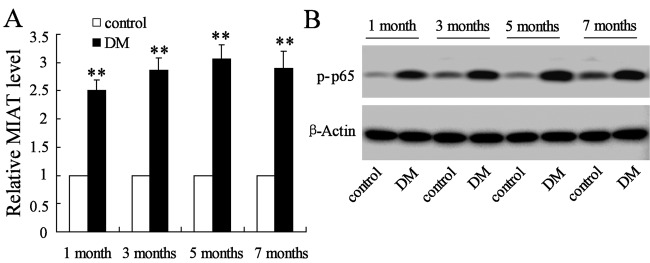
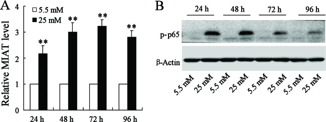
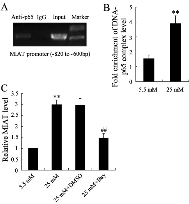
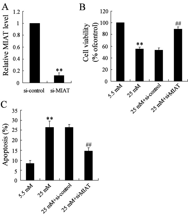
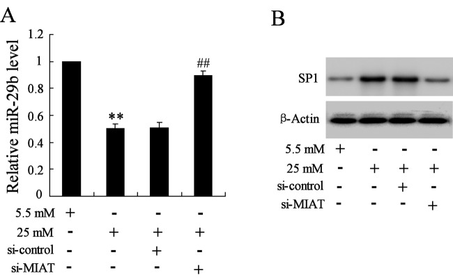
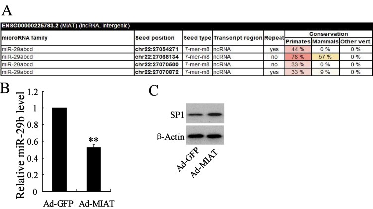
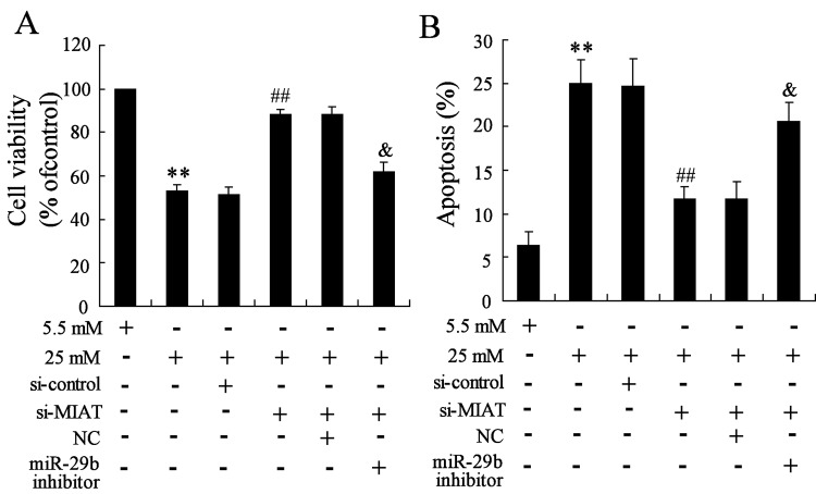
Diabetic retinopathy (DR) is a complication of diabetes mellitus (DM) and is the leading cause of vision loss globally. However, the pathogenic mechanism and clinical therapy still needs further improvement. The biologic significance of myocardial infarction associated transcript (MIAT) in DR remains unknown. Here, we aim to explore the mechanism between MIAT and DR, which is essential for RD. Streptozotocin (STZ) was used to induce DM mice and high glucose was used to stimulate cells. ChIP was used to detect the binding activity between nuclear factor κB (NF-κB) and the promoter of the MIAT gene, luciferase activity assay was used to detect the target-specific selectivity between miR-29b and MIAT. The expressions of MIAT and p-p65 were increased in STZ-induced DM mice and high glucose stimulated rat retinal Müller cells (rMC-1) cells. ChIP results revealed that high glucose promoted the binding activity between NF-κB and MIAT, while Bay11-7082 acted as an inhibitor for NF-κB that suppressed the binding activity. miR-29b controled MIAT to regulate its expression and MIAT overexpression suppressed miR-29b, but promoted Sp1. High glucose stimulation increased the cell apoptosis and decreased the cell activity, while MIAT suppression reversed the effect induced by high glucose, however, miR-29b knockdown reversed the effects induced by MIAT suppression. Our results provided evidence that the mechanism of cell apoptosis in DR might be associated with the regulation of MIAT, however, miR-29b acted as a biomarker that was regulated by MIAT and further regulated cell apoptosis in DR.

Keywords: MIAT; cell apoptosis; diabetic retinopathy; high glucose; miR-29b.


Conflict of interest statement

The authors declare that there are no competing interests associated with the manuscript.

Figures

Figure 1
Figure 1. The expression level of MIAT and p-p65 in Müller cells
(A) STZ supplementation significantly increased the expression level of MIAT than citrate buffer supplementation in mice. (B) The expression level of p-p65 was significantly increased in STZ-induced DM mice compared with citrate buffer-induced control. **P<0.01 compared with control, β-actin served as the internal control.
Figure 2
Figure 2. The effects of glucose concentration on the expression of MIAT and p-p65
Comparing with the normal glucose supplementation, the rMC-1 cells stimulated by high glucose contributed to the elevation of MIAT expression (A) and p-p65 expression (B). **P<0.01 compared with normal glucose supplementation, β-actin served as the internal control.
Figure 3
Figure 3. The relationship of NF-κB and MIAT
(A) NF-κB directly binds to MIAT promoter. (B) High glucose promoted the binding activity of NF-κB and MIAT promoter in rMC-1 cells. (C) rMC-1 cells pretreated with Bay11-7082 and then stimulated by high glucose, the expression of MIAT was significantly decreased. **P<0.01 compared with normal glucose, ##P<0.01 compared with high glucose + DMSO.
Figure 4
Figure 4. The effects of MIAT knockdown on cell viability and apoptosis in high glucose-induced rMC-1 cells
(A) The transfected efficiency was significantly decreased. (B) MIAT knockdown significantly reversed the decrease in cell viability induced by high glucose. (C) MIAT knockdown significantly reversed the increase in cell apoptosis induced by high glucose. **P<0.01 compared with normal glucose, ##P<0.01 compared with high glucose + si-control.
Figure 5
Figure 5. The effects of MIAT suppression on the expression of miR-29 and Sp1 in high glucose-induced rMC-1 cells
(A) MIAT suppression significantly reversed the decreased expression of miR-29 induced by high glucose. (B) MIAT suppression significantly reversed the increase expression of Sp1 induced by high glucose. **P<0.01 compared with normal glucose, ##P<0.01 compared with high glucose + si-control.
Figure 6
Figure 6. miR-29b targets MIAT to regulate its expression
(A) TargetScan database predicted that miR-29b has highly conserved target sequence with 3′-UTR of MIAT. MIAT overexpression dramatically decreased the expression of miR-29b (B), while increased the expression of Sp1 in rMC-1 cells (C). **P<0.01 compared with Ad-GFP, β-actin served as the internal control.
Figure 7
Figure 7. Interaction of MIAT and miR-29b on high glucose induced rMC-1 cells
(A) MIAT suppression significantly reversed the decrease in cell viability induced by high glucose, while miR-29b knockdown significantly reversed the effect induced by MIAT suppression. (B) MIAT suppression significantly reversed the increase in cell apoptosis induced by high glucose, however, miR-29b knockdown significantly reversed the effect induced by MIAT suppression. **P<0.01 compared with normal glucose, ##P<0.01 compared with high glucose + si-control, &P<0.01 compared with high glucose + si-MIAT + NC.

References

    1. Zimmet P.Z., Magliano D.J., Herman W.H. and Shaw J.E. (2014) Diabetes: a 21st century challenge. Lancet Diabetes Endocrinol. 2, 56–64 - PubMed
    1. Merovci A., Solis-Herrera C., Daniele G., Eldor R., Fiorentino T.V., Tripathy D. et al. (2014) Dapagliflozin improves muscle insulin sensitivity but enhances endogenous glucose production. J. Clin. Invest. 124, 509–514 - PMC - PubMed
    1. American Diabetes Association (2014) Diagnosis and classification of diabetes mellitus. Diabetes Care 37, S81–S90 - PubMed
    1. Bourne R.R., Jonas J.B., Flaxman S.R., Keeffe J., Leasher J., Naidoo K. et al. (2014) Prevalence and causes of vision loss in high-income countries and in Eastern and Central Europe: 1990-2010. Br. J. Ophthalmol. 98, 629–638 - PubMed
    1. Couturier A., Dupas B., Guyomard J.L. and Massin P. (2014) Surgical outcomes of florid diabetic retinopathy treated with antivascular endothelial growth factor. Retina 34, 1952–1959 - PubMed

Publication types