. 2017 Mar 3;7(1):52.
doi: 10.1038/s41598-017-00104-z.

A novel CCR2 antagonist inhibits atherogenesis in apoE deficient mice by achieving high receptor occupancy

Affiliations

A novel CCR2 antagonist inhibits atherogenesis in apoE deficient mice by achieving high receptor occupancy

Ilze Bot et al. Sci Rep. .

Abstract

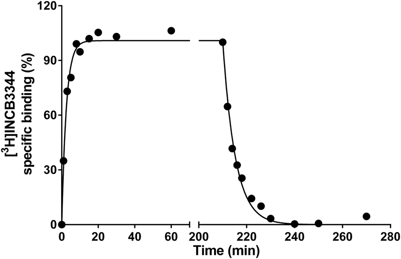
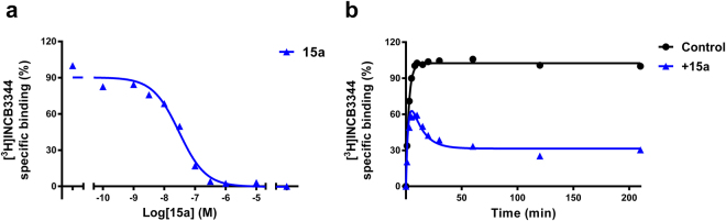
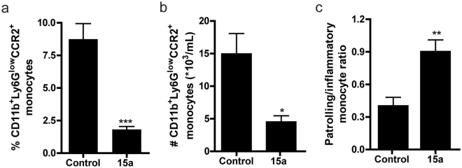
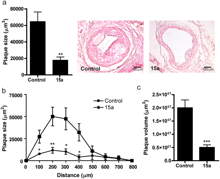
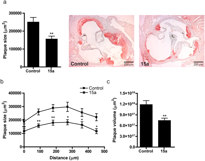
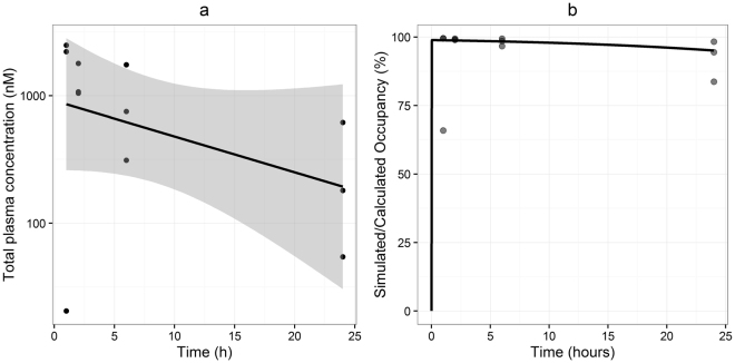
CC Chemokine Receptor 2 (CCR2) and its endogenous ligand CCL2 are involved in a number of diseases, including atherosclerosis. Several CCR2 antagonists have been developed as potential therapeutic agents, however their in vivo clinical efficacy was limited. In this report, we aimed to determine whether 15a, an antagonist with a long residence time on the human CCR2, is effective in inhibiting the development of atherosclerosis in a mouse disease model. First, radioligand binding assays were performed to determine affinity and binding kinetics of 15a on murine CCR2. To assess the in vivo efficacy, western-type diet fed apoE-/- mice were treated daily with 15a or vehicle as control. Treatment with 15a reduced the amount of circulating CCR2+ monocytes and the size of the atherosclerotic plaques in both the carotid artery and the aortic root. We then showed that the long pharmacokinetic half-life of 15a combined with the high drug concentrations ensured prolonged CCR2 occupancy. These data render 15a a promising compound for drug development and confirms high receptor occupancy as a key parameter when targeting chemokine receptors.


Conflict of interest statement

The authors declare no competing financial interests.

Figures

Figure 1
Figure 1
Chemical structure of CCR2 antagonist 15a.
Figure 2
Figure 2
Kinetic characterization of [3H]INCB3344 in murine CCR2. Association and dissociation kinetics of 5 nM [3H]INCB3344 binding to membranes of CHO cell membranes transiently expressing murine CCR2 at 25 °C. Dissociation was initiated by the addition of 10 µM INCB3344. Association and dissociation data were fitted using a monophasic exponential association, or exponential decay model, respectively. Graphs shown are representative from one experiment performed in duplicate. Data for all association and dissociation experiments (n = 3) are presented in Table 1.
Figure 3
Figure 3
Characterization of 15a in murine CCR2. (a) Displacement of [3H]INCB3344 binding from CHO cell membranes transiently expressing murine CCR2 at 25 °C, upon addition of increasing concentrations of compound 15a. (b) Competition association of 5 nM [3H]INCB3344 binding to membranes of CHO cells transiently expressing murine CCR2 at 25 °C, in the absence or presence of 30 nM 15a. Data were fitted to the competition association model of Motulsky and Mahan. Graphs shown are representative from one experiment performed in duplicate. Data for kinetic experiments (n = 3) are presented in Table 1.
Figure 4
Figure 4
Effect of 15a treatment on the amount of monocytes. (a) At 2 hours after injection and two weeks of treatment, 15a reduced the relative amount of circulating CCR2+ monocytes (defined as CD11b+Ly6Glow cells). (b) At 18 hours after the last injection (after 4 weeks of treatment), the number of circulating CD11b+Ly6GlowCCR2+ cells was significantly reduced by 15a treatment. (c) The ratio of patrolling monocytes and pro-inflammatory monocytes was significantly increased upon treatment with 15a (*P < 0.05, **P < 0.01, ***P < 0.001). Graphs shown are representative from one experiment with n = 5 per group in panel (a) and n = 6 per group in panels (b) and (c).
Figure 5
Figure 5
Inhibition of lesion development in the carotid artery by 15a. (a) The CCR2 antagonist 15a significantly reduced atherosclerotic plaque size in the carotid artery at the site of maximal stenosis. (b) Similarly, plaque volume, given as plaque size at increasing distance from the collar and (c) as total volume in µm3 was significantly reduced by 15a. Micrographs show representative images of the individual groups (100X magnification). *P < 0.05, **P < 0.01, ***P < 0.001. Graphs shown are representative from one experiment with n = 10 controls versus n = 9 15a-treated mice.
Figure 6
Figure 6
Inhibition of lesion development in the aortic root by 15a. (a) Also in the aortic root, 15a inhibited atherosclerotic lesion development after 6 weeks of Western type diet. (b) Lesion area in the aortic roots presented for each group every 90 μm of the aortic root. (c) Plaque volume was also significantly reduced in the 15a-treated group. *P < 0.05, **P < 0.01. Graphs shown are representative from one experiment with n = 10 controls versus n = 9 15a-treated mice.
Figure 7
Figure 7
Macrophage content in carotid artery and aortic root atherosclerotic lesions. (a) Macrophage content in the carotid artery lesions as measured by MOMA2 staining was reduced upon treatment with 15a, both in total macrophage positive area (left panel) and relatively as percentage of lesion size (right panel). Micrographs show representative images of the individual groups (100X magnification). (b) Also in the aortic root, 15a significantly reduced the macrophage+ area (left panel). Relative macrophage content did not significantly differ between the groups (right panel). Micrographs show representative images of the individual groups (40X magnification). *P < 0.05, **P < 0.01. Graphs shown are representative from one experiment with n = 10 controls versus n = 9 15a-treated mice.
Figure 8
Figure 8
Total plasma concentrations, calculated and simulated target occupancy. (a) Observed plasma concentrations (dots), linear fit of the data (line) and 95% confidence interval of the linear regression (shaded area). (b) Equilibrium occupancy derived from the experimentally observed plasma concentrations (dots) and simulated occupancy on basis of the observed binding kinetics and plasma elimination rate (line). Graphs shown are representative from one experiment (n = 3 per time point).

References

    1. Griffith JW, Sokol CL, Luster AD. Chemokines and chemokine receptors: positioning cells for host defense and immunity. Annu Rev Immunol. 2014;32:659–702. doi: 10.1146/annurev-immunol-032713-120145. - DOI - PubMed
    1. Viola A, Luster AD. Chemokines and their receptors: drug targets in immunity and inflammation. Annual review of pharmacology and toxicology. 2008;48:171–197. doi: 10.1146/annurev.pharmtox.48.121806.154841. - DOI - PubMed
    1. White GE, Iqbal AJ, Greaves DR. CC chemokine receptors and chronic inflammation–therapeutic opportunities and pharmacological challenges. Pharmacological reviews. 2013;65:47–89. doi: 10.1124/pr.111.005074. - DOI - PubMed
    1. Boring L, Gosling J, Cleary M, Charo IF. Decreased lesion formation in CCR2−/− mice reveals a role for chemokines in the initiation of atherosclerosis. Nature. 1998;394:894–897. doi: 10.1038/29788. - DOI - PubMed
    1. Dawson TC, Kuziel WA, Osahar TA, Maeda N. Absence of CC chemokine receptor-2 reduces atherosclerosis in apolipoprotein E-deficient mice. Atherosclerosis. 1999;143:205–211. doi: 10.1016/S0021-9150(98)00318-9. - DOI - PubMed

Publication types

MeSH terms