

Lymphocytic esophagitis: Still an enigma a decade later
- PMID: 28246468
- PMCID: PMC5311104
- DOI: 10.3748/wjg.v23.i6.949
Lymphocytic esophagitis: Still an enigma a decade later
Abstract
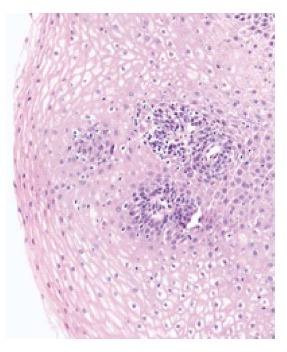
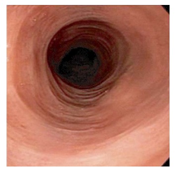
Lymphocytic esophagitis (LE) is a clinicopathologic entity first described by Rubio et al in 2006. It is defined as peripapillary intraepithelial lymphocytosis with spongiosis and few or no granulocytes on esophageal biopsy. This definition is not widely accepted and the number of lymphocytes needed to make the diagnosis varied in different studies. Multiple studies have described potential clinical associations and risk factors for LE, such as old age, female gender and smoking history. This entity was reported in inflammatory bowel disease in the pediatric population but not in adults. Other associations include gastroesophageal reflux disease and primary esophageal motility disorders. The most common symptom is dysphagia, with a normal appearing esophagus on endoscopy, though esophageal rings, webs, nodularities, furrows and strictures have been described. Multiple treatment modalities have been used such as proton pump inhibitors and topical steroids. Esophageal dilation seems to be therapeutic when dysphagia is present along with esophageal narrowing secondary to webs, rings or strictures. The natural history of the disease remains unclear and needs to be better delineated. Overall, lymphocytic esophagitis seems to have a chronic and benign course, except for two cases of esophageal perforation in the literature, thought to be secondary to this entity.
Keywords: CD4 T-cells; Lymphocytic esophagitis; dysphagia; esophageal dilation; esophageal rings; gastroesophageal reflux disease; inflammatory bowel disease; intraepithelial lymphocytes; proton pump inhibitors; spongiosis.
Conflict of interest statement
Conflict-of-interest statement: All authors have no conflict of interest.
Figures
References
-
- Rubio CA, Sjödahl K, Lagergren J. Lymphocytic esophagitis: a histologic subset of chronic esophagitis. Am J Clin Pathol. 2006;125:432–437. - PubMed
-
- Ismail-Beigi F, Horton PF, Pope CE. Histological consequences of gastroesophageal reflux in man. Gastroenterology. 1970;58:163–174. - PubMed
-
- Cucchiara S, D’Armiento F, Alfieri E, Insabato L, Minella R, De Magistris TM, Scoppa A. Intraepithelial cells with irregular nuclear contours as a marker of esophagitis in children with gastroesophageal reflux disease. Dig Dis Sci. 1995;40:2305–2311. - PubMed
-
- Wang HH, Mangano MM, Antonioli DA. Evaluation of T-lymphocytes in esophageal mucosal biopsies. Mod Pathol. 1994;7:55–58. - PubMed
Publication types
MeSH terms
Substances
LinkOut - more resources
Full Text Sources
Other Literature Sources
Medical
Research Materials
Miscellaneous
