. 2017:2017:7306192.
doi: 10.1155/2017/7306192. Epub 2017 Jan 26.

Impact of Combined Subthalamic Nucleus and Substantia Nigra Stimulation on Neuropsychiatric Symptoms in Parkinson's Disease Patients

Affiliations

Impact of Combined Subthalamic Nucleus and Substantia Nigra Stimulation on Neuropsychiatric Symptoms in Parkinson's Disease Patients

U Hidding et al. Parkinsons Dis. 2017.

Abstract

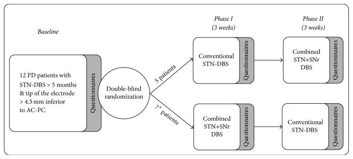
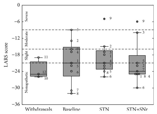
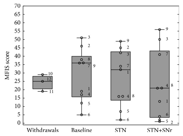
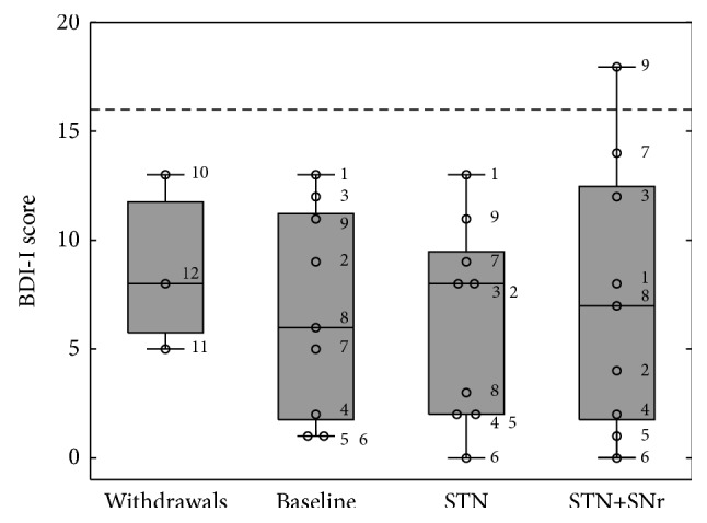
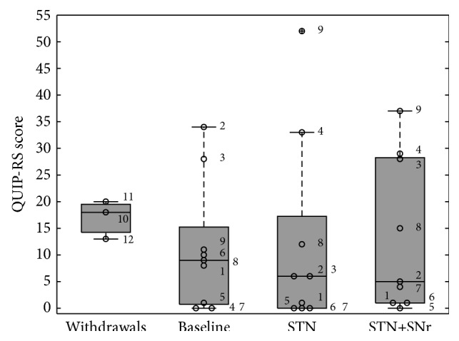
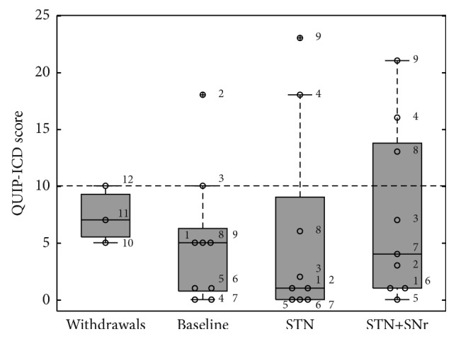
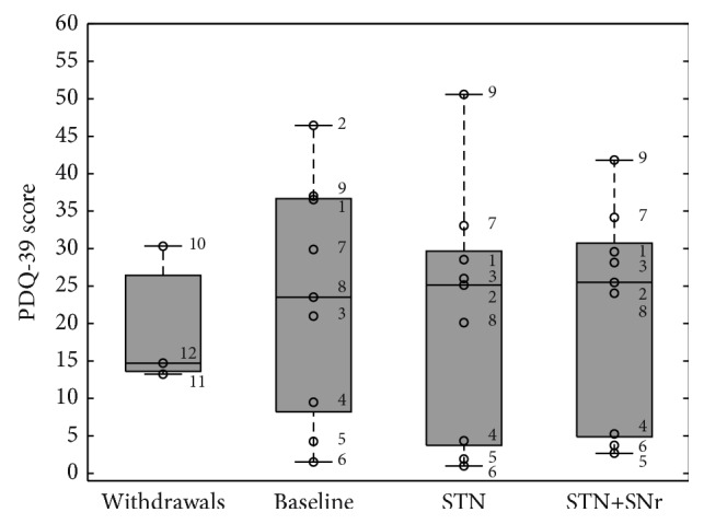
The goal of the study was to compare the tolerability and the effects of conventional subthalamic nucleus (STN) and combined subthalamic nucleus and substantia nigra (STN+SNr) high-frequency stimulation in regard to neuropsychiatric symptoms in Parkinson's disease patients. In this single center, randomized, double-blind, cross-over clinical trial, twelve patients with advanced Parkinson's disease (1 female; age: 61.3 ± 7.3 years; disease duration: 12.3 ± 5.4 years; Hoehn and Yahr stage: 2.2 ± 0.39) were included. Apathy, fatigue, depression, and impulse control disorder were assessed using a comprehensive set of standardized rating scales and questionnaires such as the Lille Apathy Rating Scale (LARS), Modified Fatigue Impact Scale (MFIS), Becks Depression Inventory (BDI-I), Questionnaire for Impulsive-Compulsive Disorders in Parkinson's Disease Rating Scale (QUIP-RS), and Parkinson's Disease Questionnaire (PDQ-39). Three patients that were initially assigned to the STN+SNr stimulation mode withdrew from the study within the first week due to discomfort. Statistical comparison of data retrieved from patients who completed the study revealed no significant differences between both stimulation conditions in terms of mean scores of scales measuring apathy, fatigue, depression, impulse control disorder, and quality of life. Individual cases showed an improvement of apathy under combined STN+SNr stimulation. In general, combined STN+SNr stimulation seems to be safe in terms of neuropsychiatric side effects, although careful patient selection and monitoring in the short-term period after changing stimulation settings are recommended.


Conflict of interest statement

The authors declare that the research was conducted in the absence of any commercial or financial relationships that could be construed as a potential conflict of interests. All authors declare no relevant conflict of interests. Some of the authors (U. Hidding, A. Gulberti, A. Horn, C. Buhmann, W. Hamel, C. K. E. Moll, and M. Pötter-Nerger) have occasionally been reimbursed for travel expenses from Medtronic Inc. C. Buhmann served on the scientific advisory board for GSK and UCB Pharma and received honoraria for lectures from GSK, Medtronic, Orion Pharma, and UCB. M. Pötter-Nerger served in advisory boards for Boston scientific, St. Jude, and AbbVie.

Figures

Figure 1
Figure 1
Simplified model of DBS induced modulation of the limbic basal ganglia loops including brainstem projections. N. caud.: nucleus caudatus; GPe: globus pallidus externus; GPi: globus pallidus internus; STN: nucleus subthalamicus; SNr: substantia nigra pars reticulata.
Figure 2
Figure 2
Study design: randomized cross-over trial over 6 weeks. After baseline assessment, PD patients were assigned in phase I to either conventional STN-DBS or combined STN+SNr stimulation. After 3 weeks, patients were switched to the other stimulation mode. () Three of the 7 patients starting the phase I with a combined STN+SNr-DBS withdrew within the first week.
Figure 3
Figure 3
Stereotactic reconstruction: squares indicate the localization of the most ventral DBS electrode contacts, superimposed on frontal sections of the stereotactic atlas of Morel, at a level 5 mm behind midcommissural point [50]. Circles indicate the ventral STN border as defined by standard electrophysiological criteria. Diamonds indicate the presence of unambiguous SNr cell activity on MER tracks. Colors represent individual patients. The dashed red lines denote midline and AC-PC level, respectively. STN: subthalamic nucleus; SNr: substantia nigra pars reticulata; SNc: substantia nigra pars compacta; CP: cerebral peduncle; RN, red nucleus.
Figure 4
Figure 4
Boxplots of LARS scores: the box plot depicted on the left represents the baseline values of the three patients who withdrew. The other three box plots represent the values of the 9 PD patients who have completed the study at the three experimental time points: (1) baseline, (2) STN alone, and (3) combined STN+SNr-DBS. The numbered circles represent the single patients as listed in Table 1. The dashed horizontal lines represent the cut-offs as previously described.
Figure 5
Figure 5
Boxplots of MFIS scores: the box plot depicted on the left represents the baseline values of the three patients who withdrew. The other three box plots represent the values of the 9 PD patients who have completed the study at the three experimental time points: (1) baseline, (2) STN alone, and (3) combined STN+SNr-DBS. The numbered circles represent the single patients as listed in Table 1.
Figure 6
Figure 6
Boxplots of BDI-I scores: the box plot depicted on the left represents the baseline values of the three patients who withdrew. The other three box plots represent the values of the 9 PD patients who have completed the study at the three experimental time points: (1) baseline, (2) STN alone, and (3) combined STN+SNr-DBS. The numbered circles represent the single patients as listed in Table 1. The dashed horizontal line represents the cut-off for PD patients as previously discussed in Material and Methods.
Figure 7
Figure 7
Boxplots of QUIP-RS scores: the box plot depicted on the left represents the baseline values of the three patients who withdrew. The other three box plots represent the values of the 9 PD patients who have completed the study at the three visits: (1) baseline, (2) STN alone, and (3) combined STN+SNr-DBS. The numbered circles represent the single patients as listed in Table 1. A cut-off for the total QUIP-RS score is not available.
Figure 8
Figure 8
Boxplots of combined QUIP-ICD scores: the box plot depicted on the left represents the baseline values of the three patients who withdrew. The other three box plots represent the values of the 9 PD patients who have completed the study at the three experimental time points: (1) baseline, (2) STN alone, and (3) combined STN+SNr-DBS. The numbered circles represent the single patients as listed in Table 1. For the combined QUIP-ICD score the cut-off point is ≥10.
Figure 9
Figure 9
Boxplots of PDQ-39 scores expressed as Parkinson's disease summary index (PDSI) scores: the box plot depicted on the left represents the baseline values of the three patients who withdrew. The other three box plots represent the values of the 9 PD patients who have completed the study at the three experimental time points: (1) baseline, (2) STN alone, and (3) combined STN+SNr-DBS. The numbered circles represent the single patients as listed in Table 1.

Similar articles

Cited by

References

    1. Castrioto A., Thobois S., Carnicella S., Maillet A., Krack P. Emotional manifestations of PD: neurobiological basis. Movement Disorders. 2016;31(8):1103–1113. doi: 10.1002/mds.26587. - DOI - PubMed
    1. Martinez-Fernandez R., Pelissier P., Quesada J.-L., et al. Postoperative apathy can neutralise benefits in quality of life after subthalamic stimulation for Parkinson's disease. Journal of Neurology, Neurosurgery and Psychiatry. 2016;87(3):311–318. doi: 10.1136/jnnp-2014-310189. - DOI - PubMed
    1. Sierra M., Carnicella S., Strafella A. P., et al. Apathy and impulse control disorders: yin & yang of dopamine dependent behaviors. Journal of Parkinson's Disease. 2015;5(3):625–636. doi: 10.3233/jpd-150535. - DOI - PubMed
    1. Weintraub D., Burn D. J. Parkinson's disease: the quintessential neuropsychiatric disorder. Movement Disorders. 2011;26(6):1022–1031. doi: 10.1002/mds.23664. - DOI - PMC - PubMed
    1. Pont-Sunyer C., Hotter A., Gaig C., et al. The onset of nonmotor symptoms in parkinson's disease (The ONSET PD Study) Movement Disorders. 2015;30(2):229–237. doi: 10.1002/mds.26077. - DOI - PubMed

LinkOut - more resources