Review
. 2017:59:175-200.
doi: 10.1007/978-3-319-44820-6_6.

Translational Control of Germ Cell Decisions

Affiliations
Review

Translational Control of Germ Cell Decisions

Kumari Pushpa et al. Results Probl Cell Differ. 2017.

Abstract

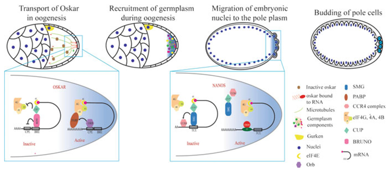
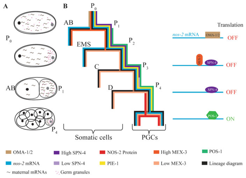
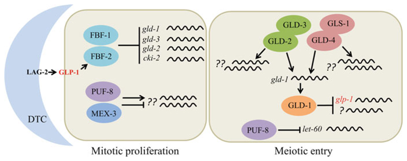
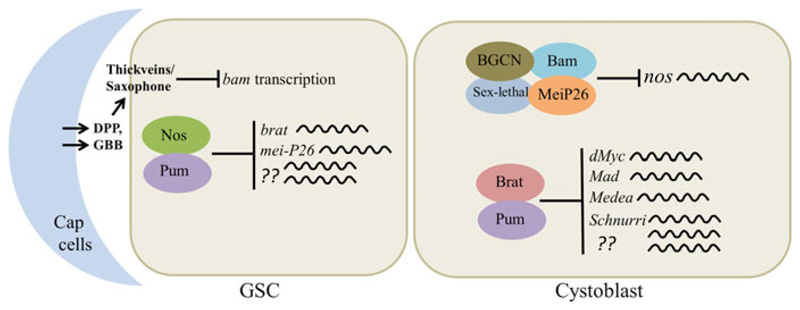
Germline poses unique challenges to gene expression control at the transcriptional level. While the embryonic germline maintains a global hold on new mRNA transcription, the female adult germline produces transcripts that are not translated into proteins until embryogenesis of subsequent generation. As a consequence, translational control plays a central role in governing various germ cell decisions including the formation of primordial germ cells, self-renewal/differentiation decisions in the adult germline, onset of gametogenesis and oocyte maturation. Mechanistically, several common themes such as asymmetric localization of mRNAs, conserved RNA-binding proteins that control translation by 3' UTR binding, translational activation by the cytoplasmic elongation of the polyA tail and the assembly of mRNA-protein complexes called mRNPs have emerged from the studies on Caenorhabditis elegans, Xenopus and Drosophila. How mRNPs assemble, what influences their dynamics, and how a particular 3' UTR-binding protein turns on the translation of certain mRNAs while turning off other mRNAs at the same time and space are key challenges for future work.


Figures

Fig. 6.1
Fig. 6.1
Translational control during germ cell specification in Drosophila. Germplasm is assembled during oogenesis by Oskar. oskar mRNA is transported in an inactive form and deposited at the posterior pole where its translation is activated. oskar translation is suppressed by Bruno with the help of Cup. At the posterior, oskar translation is activated by ORB; ORB recruits PABP and facilitates translational activation. Oskar further recruits other germplasm components during late stages of oogenesis. The early Drosophila embryo is a syncytium; at the start of cellularization, some of the posterior nuclei and the surrounding germplasm form the pole cells (PGCs) by budding. nanos mRNA is translationally suppressed in the anterior region by Smaug (Smg) which recruits Cup and prevents translation initiation. At the posterior end, nos mRNA is bound by Oskar, which prevents Smg from binding to nos-2 3′ UTR, which derepresses nos mRNA. Smaller cells diagrammed in the top left two cartoons represent the nurse cells
Fig. 6.2
Fig. 6.2
Translational control during germ cell specification in C. elegans. (a) Schematic representation of asymmetric cleavage and asymmetric distribution of maternal components. Pink dots and tilde-like structures represent the germ granules and the maternal mRNAs, respectively. (b) Distribution patterns of RBPs and nos-2 mRNA during early embryonic cleavages; colors representing the different components are indicated at the bottom. Translation of nos-2 mRNA is suppressed sequentially by OMA-1 and OMA-2 in oocytes (not shown here), by MEX-3 in the AB blastomere, and by SPN-4 in the P lineage until P3. The rapid decrease in the SPN-4 to POS-1 ratio in P4 enables POS-1 to compete out SPN-4 for binding to the nos-2 3′ UTR, which depresses nos-2 translation in P4
Fig. 6.3
Fig. 6.3
Translational control of the mitosis–meiosis decision in C. elegans. The LAG-2 ligand produced by the somatic cell called the distal tip cell (DTC) activates the GLP-1 receptor present on germ cells. This results in the transcriptional activation of the RBP FBF-2, which along with FBF-1 inhibits meiotic entry by suppressing the translation of gld-1, gld-3, gld-2, and cki-2 mRNAs. The RBPs PUF-8 and MEX-3 promote proliferation, possibly by regulating the translation of unknown mRNAs. Although a single proliferating cell is shown in this cartoon, the mitotic region extends to about 20-cell diameters from DTC. The entire proliferative zone comprises of a total of ~200 cells. The schematic on the right represents a cell from the transition zone. In the transition zone, GLP-1 activity and the levels of FBFs decrease, resulting in the expression of FBF targets such as GLD-1 and GLD-2. GLD-1 represses the translation of glp-1 and unknown mRNAs to promote meiotic entry. GLD-2 and GLD-4 (PAPs) promote GLD-1 expression. In addition, these two PAPs promote meiotic entry independently of GLD-1 by regulating the translation of unknown mRNAs. Furthermore, PUF-8 facilitates meiotic entry by repressing the translation of let-60, which encodes RAS, a well-known proliferation-promoting factor, in this zone
Fig. 6.4
Fig. 6.4
Translational control of the self-renewal/differentiation decision in the Drosophila ovary. In the Drosophila ovary, signaling from the niche (cap cells) suppresses bam transcription in the GSC. Apart from this, Nos-Pum pair represses the translation of brat, mei-P26, and other unknown mRNAs to prevent premature differentiation. The GSC divides such that one daughter cell is oriented away from the niche and does not receive sufficient niche signals to suppress bam. Bam forms a multi-protein complex with Mei-P26, Sex-lethal, and BGCN and suppresses nos translation. Absence of Nos derepresses the brat mRNA leading to Brat expression, which partners with Pum and inhibits the translation of dMyc, Mad, Medea, and Schnurri to promote differentiation
Fig. 6.5
Fig. 6.5
Nanos mRNP is crucial for spermatogonial stem cell maintenance in mice. In mouse, GDNF and FGF9 secreted by Sertoli cells activate Nanos2 expression in SSCs. Nanos2 sequesters differentiationpromoting mRNAs into mRNPs along with other RBPs
Fig. 6.6
Fig. 6.6
Translational regulation during meiotic progression, oocyte growth, and maturation in C. elegans. A cartoon of the adult hermaphrodite gonad is shown at the top. Orientation: distal to the left and proximal to the right. C. elegans gonad with different regions marked. Horizontal bars indicate expression patterns of the RBPs that regulate meiotic progression and/or oocyte maturation. Intensity variations of the color reflect the concentrations of the corresponding proteins. The RBPs and their corresponding target mRNAs in the different stages of meiotic development are shown at the bottom. See text for a more detailed description
Fig. 6.7
Fig. 6.7
Sequential activation of polyadenylation during oocyte maturation in Xenopus. (a) Outline of the transcriptional and translational status at the key stages of oocyte maturation. (b) Summary of the signaling cascade activated by progesterone during oocyte maturation. Progesterone stimulation releases Ringo mRNA from Pum2-mediated repression. Ringo associates with Cdc-2, phosphorylates CPEB, Cdc-2, and Musashi. Phosphorylated Musashi recruits the PAP Gld2 and activates translation of early class mRNAs. One of them is Mos mRNA. Mos activates the MAPK pathway by phosphorylating MEK. MAPK in turn phosphorylates CPEB at a site distinct from the one phosphorylated by Ringo/cyclin B-Cdc-2. (c) Phosphorylation of CPEB activates translation of masked mRNAs. Translationally inactive (masked) mRNAs are bound by CPEB, ePAB, PARN, Gld-2, and Maskin. Maskin binds to CPEB and eIF4E, blocking association of eIF4E with eIF4G. Polyadenylation–deadenylation cycles by GLD2 and PARN keep the polyA tail short. Phosphorylation of CPEB expels PARN, leading to polyA tail extension. Another factor released from the complex is ePAB, which now binds to the polyA tail and associates with eIF4E by competing out Maskin and recruits eIF4G leading to translation activation

References

    1. Ariz M, Mainpal R, Subramaniam K. C. elegans RNA-binding proteins PUF-8 and MEX-3 function redundantly to promote germline stem cell mitosis. Dev Biol. 2009;326:295–304. - PMC - PubMed
    1. Arumugam K, Macnicol MC, Macnicol AM. Autoregulation of Musashi1 mRNA translation during Xenopus oocyte maturation. Mol Reprod Dev. 2012a;79:553–563. - PMC - PubMed
    1. Arumugam K, MacNicol MC, Wang Y, Cragle CE, Tackett AJ, Hardy LL, MacNicol AM. Ringo/cyclin-dependent kinase and mitogen-activated protein kinase signaling pathways regulate the activity of the cell fate determinant Musashi to promote cell cycle re-entry in Xenopus oocytes. J Biol Chem. 2012b;287:10639–10649. - PMC - PubMed
    1. Austin J, Kimble J. glp-1 is required in the germ line for regulation of the decision between mitosis and meiosis in C. elegans. Cell. 1987;51:589–599. - PubMed
    1. Barnard DC, Ryan K, Manley JL, Richter JD. Symplekin and xGLD-2 are required for CPEB-mediated cytoplasmic polyadenylation. Cell. 2004;119:641–651. - PubMed

Publication types

LinkOut - more resources