

Update from the 4th Edition of the World Health Organization Classification of Head and Neck Tumours: Mucosal Melanomas
- PMID: 28247222
- PMCID: PMC5340730
- DOI: 10.1007/s12105-017-0789-y
Update from the 4th Edition of the World Health Organization Classification of Head and Neck Tumours: Mucosal Melanomas
Abstract
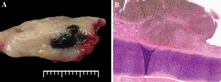
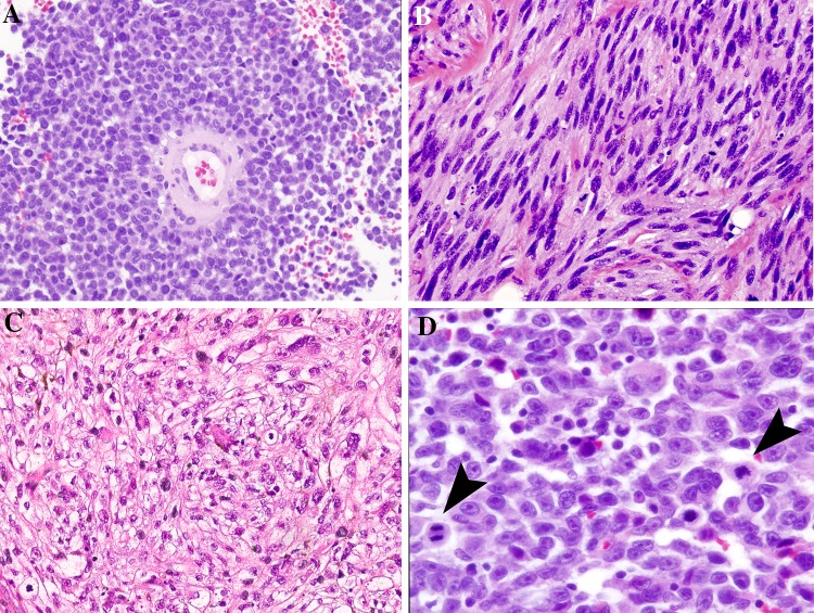
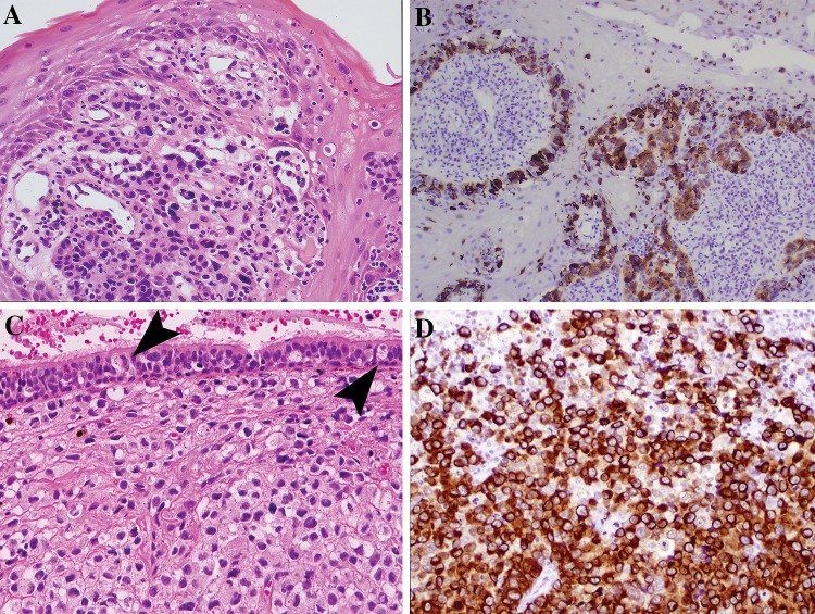
The updated edition of The World Health Organization Classification of Tumours of the Head and Neck includes discussions on mucosal melanoma of both the sinonasal and oral cavity. Since the prior edition, sinonasal origin is now recognized as the most common site of occurrence of mucosal melanoma in the head and neck (66%) with oral cavity representing 25% of cases. Histologic features of mucosal melanomas vary widely from spindled, epithelioid, and pleomorphic to rhabdoid, plasmacytoid and undifferentiated. Additionally, mucosal melanomas are commonly amelanotic (or minimal pigmentation) (~50%) leading to overlapping features and diagnostic challenges in differentiating mucosal melanomas from other small cell/undifferentiated sinonasal tumors. Since the last edition, formal staging of head and neck mucosal melanomas was added to the American Joint Committee on Cancer entities, though the traditional histologic features that have prognostic significance in cutaneous melanomas fail to stratify mucosal melanomas (i.e. tumor thickness, ulceration). Interestingly, while melanomas of all sites are a malignancy derived from melanocytes, mucosal melanomas are now recognized to have distinct molecular alterations compared to cutaneous or uveal melanomas. BRAF V600E mutations are rare (<6%) in mucosally derived melanomas compared to a rate of 50% in cutaneous melanomas. CD117 (C-Kit) mutations are the most common alteration encountered (~25%) in mucosal sites with potential therapeutic targetability. The recognition of the distinct genetic changes in this subgroup of melanomas means that therapy advances in cutaneous melanomas may not translate to head and neck mucosal melanomas and clinical trials specific to this subgroup of patients are needed.
Keywords: CD117 mutations; Mucosal melanoma; Oral melanoma; Sinonasal melanoma.
Conflict of interest statement
Conflict of interest
The author has no conflict of interest to disclose.
Research Involving Animal and Human Rights
This article does not contain any studies with human participants or animals performed by the author.
Figures
References
-
- Griewank KG, Murali R, Puig-Butille JA, Schilling B, Livingstone E, Potrony M, Carrera C, Schimming T, Möller I, Schwamborn M, Sucker A, Hillen U, Badenas C, Malvehy J, Zimmer L, Scherag A, Puig S, Schadendorf D. TERT promoter mutation status as an independent prognostic factor in cutaneous melanoma. J Natl Cancer Inst. 2014;106(9). - PMC - PubMed
MeSH terms
LinkOut - more resources
Full Text Sources
Other Literature Sources
Medical
Molecular Biology Databases
Research Materials
