. 2017 Mar;11(1):55-67.
doi: 10.1007/s12105-017-0795-0. Epub 2017 Feb 28.

Update from the 4th Edition of the World Health Organization Classification of Head and Neck Tumours: Tumors of the Salivary Gland

Affiliations

Update from the 4th Edition of the World Health Organization Classification of Head and Neck Tumours: Tumors of the Salivary Gland

Raja R Seethala et al. Head Neck Pathol. 2017 Mar.

Abstract

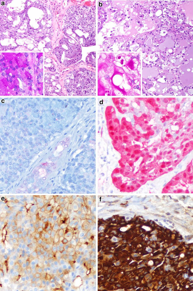
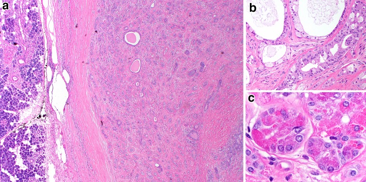
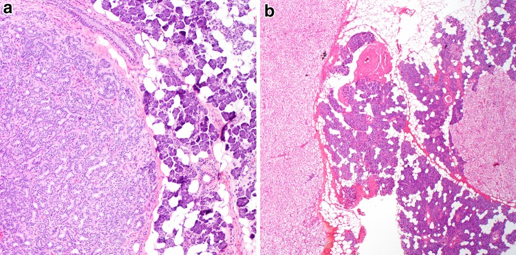
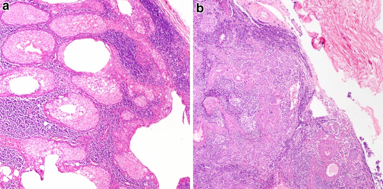
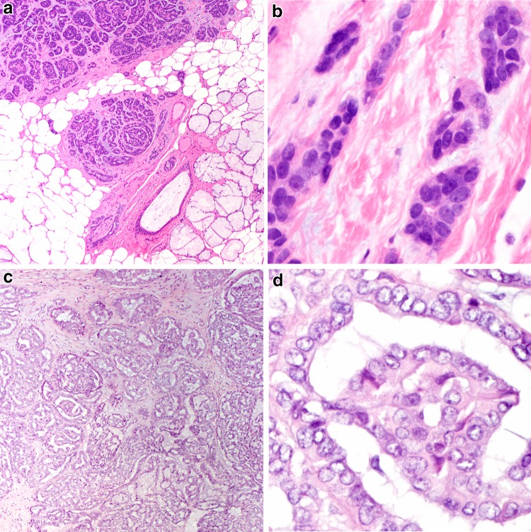
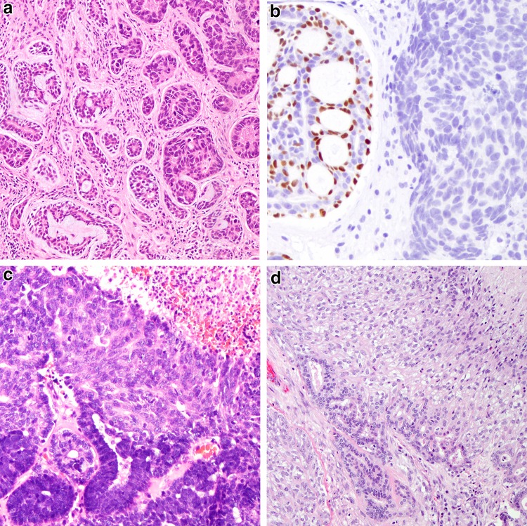
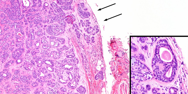
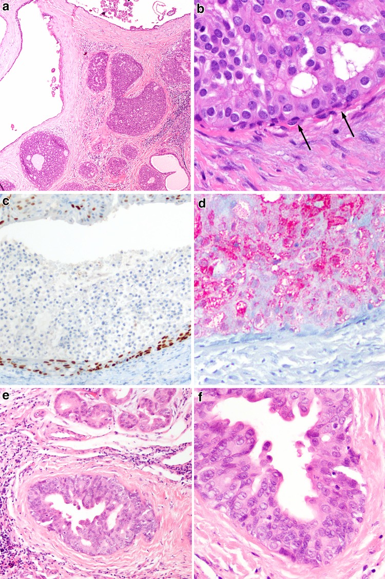
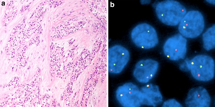
The salivary gland section in the 4th edition of the World Health Organization classification of head and neck tumors features the description and inclusion of several entities, the most significant of which is represented by (mammary analogue) secretory carcinoma. This entity was extracted mainly from acinic cell carcinoma based on recapitulation of breast secretory carcinoma and a shared ETV6-NTRK3 gene fusion. Also new is the subsection of "Other epithelial lesions," for which key entities include sclerosing polycystic adenosis and intercalated duct hyperplasia. Many entities have been compressed into their broader categories given clinical and morphologic similarities, or transitioned to a different grouping as was the case with low-grade cribriform cystadenocarcinoma reclassified as intraductal carcinoma (with the applied qualifier of low-grade). Specific grade has been removed from the names of the salivary gland entities such as polymorphous adenocarcinoma, providing pathologists flexibility in assigning grade and allowing for recognition of a broader spectrum within an entity. Cribriform adenocarcinoma of (minor) salivary gland origin continues to be divisive in terms of whether it should be recognized as a distinct category. This chapter also features new key concepts such as high-grade transformation. The new paradigm of translocations and gene fusions being common in salivary gland tumors is featured heavily in this chapter.

Keywords: Classification; Correlative; Molecular; Neoplasia; Salivary gland; Translocations; World Health Organization.


Conflict of interest statement

Conflict of interest

The authors have no financial or other conflicts of interest to report.

Ethical Approval

This article does not contain any studies with human participants or animals performed by any of the authors.

Figures

Fig. 1
Fig. 1
A comparison of acinic cell carcinoma and (mammary analogue) secretory carcinoma. a This acinic cell carcinoma shows a solid to follicular patterned proliferation of lightly basophilic acinar type cells (H&E, 100x). Inset: PAS after diastase demonstrates a granular staining in keeping with zymogen granules (H&E, 400x). b This secretory carcinoma shows a follicular pattern but with eosinophilic vacuolated cells and mucinous luminal secretions (H&E, 100x). Inset: PAS after diastase highlights globular and luminal staining in keeping with mucin (H&E, 400x). c This acinic cell carcinoma is S100 negative (200x), while the d secretory carcinoma is S100 positive (200x). e Acinic cell carcinomas show consistent apical and membranous DOG-1 staining (200x). f Secretory carcinomas typically show strong mammaglobin staining (200x)
Fig. 2
Fig. 2
Sclerosing polycystic adenosis. a This lesion consists of a tubular to cystic proliferation of glands embedded in a highly sclerotic stroma (H&E, 20x). b Apocrine and secretory areas with histiocytes are typical (H&E, 100x), as are c acinar areas with abnormal red, coarse zymogen granules (H&E, 400x)
Fig. 3
Fig. 3
Select “other epithelial lesions.” a Intercalated duct lesion demonstrating a rounded well circumscribed proliferation of tubules with the same caliber as intercalated ducts. Such lesions are designated as “intercalated duct adenomas.” (H&E, 40x). b Clear cell oncocytosis demonstrating two well demarcated nodules (H&E, 20x). This lesion is reminiscent of metastatic renal cell carcinoma but is devoid of the vascularity and hemorrhage typical of this
Fig. 4
Fig. 4
Lymphadenomas. a Sebaceous lymphadenomas consist of a well circumscribed solid and cystic proliferation of squamous and sebaceous elements (H&E, 100x). Similarly, non-sebaceous adenomas are also will demarcated with prominent lymphoid stroma. b They also demonstrate trabeculae of basaloid squamoid elements and may be variably microcystic, with the main distinction being the absence of sebaceous elements (H&E, 100x)
Fig. 5
Fig. 5
Polymorphous adenocarcinoma and cribriform adenocarcinoma of (minor) salivary gland. a Classic polymorphous adenocarcinoma, low-grade, demonstrates an infiltrative, neurotropic or targetoid pattern composed of tubulofascicular growth (H&E, 40x). b Tumor cells are phenotypically monomorphic and ductal, and composed of cells with ovoid vesicular nuclei with slight membrane irregularities and inconspicuous nuclei (H&E, 200x). c Cribriform adenocarcinoma of minor salivary gland actually consists of papillary glomeruloid structures as well as cribriform spaces (H&E, 40x). d Tumor cells are similarly monomorphic and ductal, but contain slightly larger nuclei and more markedly cleared chromatin remincent of (classic) papillary thyroid carcinoma nuclei
Fig. 6
Fig. 6
High-grade transformation in carcinomas. a Adenoid cystic carcinoma with high-grade transformation showing a transition from a more typical cribriform growth pattern consisting of angulated, hyperchromatic but monomorphic nuclei (left) to a markedly pleomorphic adenocarcinoma (right) (H&E, 100x). b A hallmark of transformation of biphasic carcinomas like adenoid cystic carcinoma is ductal overgrowth and loss of biphasic cell composition as demonstrated by the loss of p63 staining in the transformed component (right) (200x). c Acinic cell carcinoma with high-grade transformation demonstrating transition from conventional acinic cell carcinoma (bottom) with deeply basophilic zymogen granules to a solid carcinoma with necrosis (H&E, 100x). d Epithelial myoepithelial carcinoma with high-grade transformation of myoepithelial cell component as demonstrated by the overgrowth of the abluminal clear cell component, pleomorphism and necrosis (top right) (H&E, 100x)
Fig. 7
Fig. 7
Minimally invasive carcinoma ex pleomorphic adenoma. a This tumor shows very limited i.e. <1.5 mm, extension beyond its capsule (arrows) (H&E, 40x). However, even extending the cut-off to 4–6 mm appears to still correlate with favorable outcome. b Tumors with minimal invasion may be difficult to subtype given the limited carcinomatous component, but here the biphasic appearance consisting of luminal eosinophilic ductal cells and clear abluminal myoepithelial cells justifies designation as epithelial-myoepithelial carcinoma
Fig. 8
Fig. 8
Intraductal carcinoma. a Intraductal carcinoma, low-grade demonstrating a typical cystic to solid/cribriform proliferation of cells reminiscent of low-grade ductal carcinoma in situ of breast (H&E, 40x). b Cells are monomorphic and demonstrate variable eosinophilic cytoplasm (H&E, 400x), and delimited by an attenuated basal/myoepithelial cell layer (arrows). c This layer can be readily demonstrated by a p63 immunostain (100x). d The actual lesional cells are S100 positive (100x). e In contrast, intraductal carcinoma, high-grade, demonstrates an appearance more reminiscent of salivary duct carcinoma. Cancerization of acini (top) is not uncommon (H&E, 100x). f The nuclear size variation, eosinophilic cytoplasm and abortive decapitation secretions are in keeping with an apocrine phenotype akin to salivary duct carcinoma (H&E, 200x)
Fig. 9
Fig. 9
(Hyalinizing) clear cell carcinoma. a This is a clear cell tumor composed of monomorphic nests and cords of cells embedded in a hyalinized stroma (H&E, 100x). The peritumoral stroma is hyalinized while the intervening stroma is fibrocellular. b This tumor is characterized by an EWSR1-ATF1 fusion demonstratable by EWSR1 break-apart fluorescence in situ hybridization (separated red and green signals)

References

    1. Eveson JW, Auclair PL, Gnepp DR, El-Naggar AK. Tumours of the salivary gland. In: Barnes L, Eveson JW, Reichart P, Sidransky D, editors. World Health Organization classification of tumours: pathology and genetics of head and neck tumours. Lyon: IARC; 2005. p. 164.
    1. Stenman G. Fusion oncogenes in salivary gland tumors: molecular and clinical consequences. Head Neck Pathol. 2013;7(Suppl 1):S12–S19. doi: 10.1007/s12105-013-0462-z. - DOI - PMC - PubMed
    1. Chiosea SI, Griffith C, Assaad A, Seethala RR. The profile of acinic cell carcinoma after recognition of mammary analog secretory carcinoma. Am J Surg Pathol. 2012;36(3):343–350. doi: 10.1097/PAS.0b013e318242a5b0. - DOI - PubMed
    1. Skalova A, Vanecek T, Sima R, Laco J, Weinreb I, Perez-Ordonez B, et al. Mammary analogue secretory carcinoma of salivary glands, containing the ETV6-NTRK3 fusion gene: a hitherto undescribed salivary gland tumor entity. Am J Surg Pathol. 2010;34(5):599–608. - PubMed
    1. Bishop JA, Yonescu R, Batista D, Eisele DW, Westra WH. Most nonparotid “acinic cell carcinomas” represent mammary analog secretory carcinomas. Am J Surg Pathol. 2013;37(7):1053–1057. doi: 10.1097/PAS.0b013e3182841554. - DOI - PMC - PubMed

MeSH terms

LinkOut - more resources