Review
. 2014 Nov 12;1(1):41-54.
doi: 10.1016/j.jcmgh.2014.10.001. eCollection 2015 Jan.

The Barrett's Gland in Phenotype Space

Affiliations
Review

The Barrett's Gland in Phenotype Space

Stuart A C McDonald et al. Cell Mol Gastroenterol Hepatol. .

Abstract

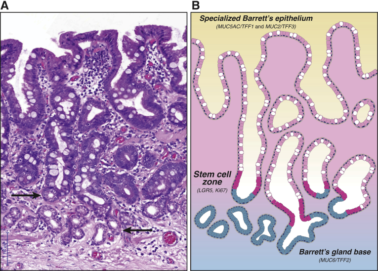
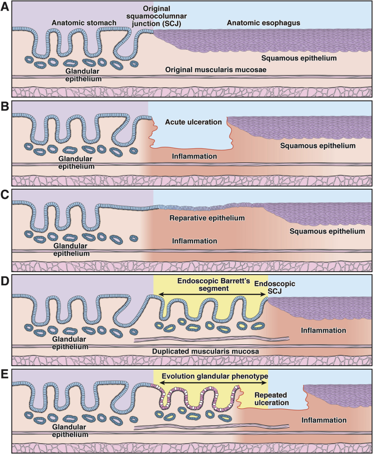
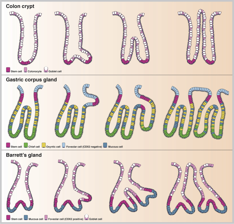
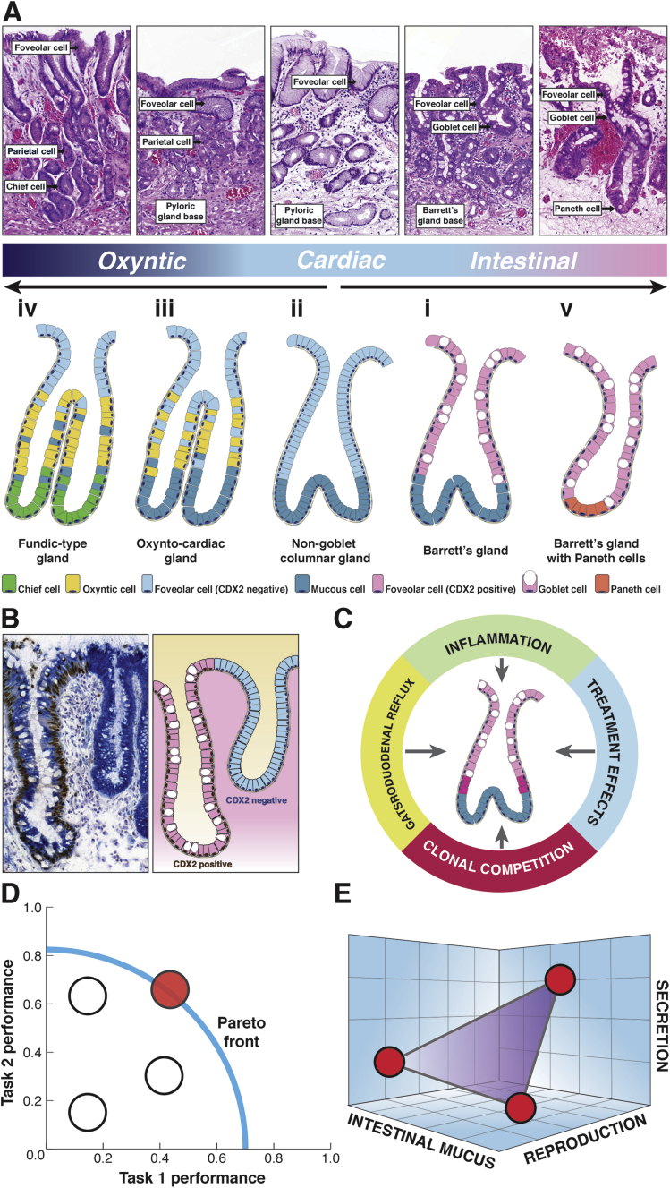
Barrett's esophagus is characterized by the erosive replacement of esophageal squamous epithelium by a range of metaplastic glandular phenotypes. These glandular phenotypes likely change over time, and their distribution varies along the Barrett's segment. Although much recent work has addressed Barrett's esophagus from the genomic viewpoint-its genotype space-the fact that the phenotype of Barrett's esophagus is nonstatic points to conversion between phenotypes and suggests that Barrett's esophagus also exists in phenotype space. Here we explore this latter concept, investigating the scope of glandular phenotypes in Barrett's esophagus and how they exist in physical and temporal space as well as their evolution and their life history. We conclude that individual Barrett's glands are clonal units; because of this important fact, we propose that it is the Barrett's gland that is the unit of selection in phenotypic and indeed neoplastic progression. Transition between metaplastic phenotypes may be governed by neutral drift akin to niche turnover in normal and dysplastic niches. In consequence, the phenotype of Barrett's glands assumes considerable importance, and we make a strong plea for the integration of the Barrett's gland in both genotype and phenotype space in future work.

Keywords: Barrett’s Esophagus; CCO, cytochrome c oxidase; Metaplasia; Neutral Drift.


Figures

Figure 1
Figure 1
Barrett’s glands show functional compartmentalization.(A) H&E photomicrograph of a series of nondysplastic Barrett’s glands (of canonical, specialized type) demonstrating abundant goblet cells. Note the mucous glands arranged as small acini at the base of these Barrett’s gland (arrows). (B) Illustration of the Barrett’s glands shown in A. The stem cell zone (shown in magenta) demonstrates maximal Ki-67 proliferative activity and LGR5 labeling on in situ hybridization. This proliferative compartment is located about one-third up the glandular axis. Labeling studies demonstrate bidirectional flow from this stem cell compartment. Specifically, specialized epithelium (a combination of MUC5AC+/TFF1+ foveolar cells and MUC2+/TFF3+ goblet cells, both shown in pink) migrates toward the luminal surface, while MUC6+/TFF2+ mucous cells (shown in blue) migrate toward the glandular base at a much slower rate. This functional compartmentalization replicates pyloric-type gland organization.
Figure 2
Figure 2
The Barrett’s gland as a unit of selection. This diagram shows the evolution of the glandular Barrett’s phenotype over time in the context of recurrent erosive esophagitis. There are various time points within the evolution of a Barrett’s segment when selection is important: during the establishment of the segment, during its progression to a stable state, and, where it does occur, during the development of dysplasia and carcinoma. At each of these time points, different traits may be under selection. (A) At baseline, the endoscopic squamocolumnar junction coincides with the anatomic gastroesophageal junction. (B) After chronic gastroduodenal reflux, the squamous lining of the esophagus is repeatedly damaged and eventually ulcerates. (C) A breach in the epithelial lining is followed by a stereotypical wound-healing response stimulated by inflammation. In the acute, proliferative stage of wound healing, competition may be dominated by selection for secretory and proliferative traits favoring (mucosal) repair. (D) Repetitive injury leads to cephalad expansion of the glandular lining within the tubular esophagus. As can be seen in other regions of the intestinal tract, remodeling is accompanied by the deposition of a complete mucosal unit, including a newly derived muscularis mucosae. (E) After the initial proliferative and remodeling stage, this simple glandular phenotype may evolve to a complex, specialized glandular phenotype.
Figure 3
Figure 3
Clonal expansion of glandular units in the gastrointestinal tract. Glandular units in the gastrointestinal tract expand as clonal patches through a fission process. Crypts in the normal colon fission on average about every 30 years, forming a small clonal patch. Fission is initiated from the stem cell zone at the crypt base (in magenta), after which the crypt unzips toward the luminal surface (top panel). Glands in the stomach fission by forming a bud from the stem cell zone at the level of the neck (in magenta) through localized cell division. The new gland grows down toward the level of the muscularis mucosae (middle panel). We propose that Barrett’s glands form clonal patches in a similar manner by forming a glandular bud at the level of the stem cell zone (again in magenta), which is located just above the MUC6/TFF2+ Barrett’s gland base. This bud extends and unzips toward the luminal surface (bottom panel). Constituent cell types of each glandular unit are indicated per panel.
Figure 4
Figure 4
The Barrett’s gland in phenotype space.(A) Morphologic definition of the spectrum of gland phenotypes found in Barrett’s esophagus. The top row shows H&E photomicrographs of the various gland phenotypes, and the second row shows an illustration of these glandular phenotypes (see the article for details). Esophageal derivation (submucosal esophageal glands, double muscularis mucosae) was verified for all examples shown here. The histology panels show labels denoting the differentiated lineage that (often in combination) defines the specific metaplasia. (Further details on immunohistochemical markers for these various differentiated lineages along with references are provided in Table 1.) The glands are numbered in order of appearance in the main text. (i) The canonical goblet-containing Barrett gland is discussed in detail in Figure 1. (ii) The non-goblet columnar gland, or cardiac gland, closely parallels the makeup and functional organization of mature Barrett’s glands, save for the absence of MUC2+ goblet cells. Foveolar cells are shown in this case in light blue because they lack CDX2 expression (see Figure 2B). (iii) The oxyntocardiac gland demonstrates parietal cell differentiation (in yellow) and is functionally equivalent to the pyloric gland. (iv) The fundic-type gland shows chief cell differentiation (light green) and is functionally equivalent to the glandular phenotype of the gastric corpus. (v) Barrett’s glands showing mature intestinal differentiation. Barrett’s glands may show Paneth cell (orange) and enterocyte differentiation (not shown), the latter possibly indicating further intestinal maturation. (B) Frozen Barrett’s material costained for Muc5AC (cytoplasmic labeling, blue substrate) and CDX2 (nuclear labeling, brown substrate). Shown are two neighboring glandular units; the left shows clear goblet cell differentiation and CDX2 expression that are both absent in the right gland (type ii and type i in Figure 2A, respectively). Foveolar differentiation and MUC5AC expression are found in both glands, showing that foveolar maturation is compatible with CDX2 expression. (C) Barrett’s glands evolve genotypically and phenotypically under the continuous Malthusian selection pressures of gastroduodenal reflux, chronic and acute inflammation, clonal competition, and finally treatment effects. (D) Schematic view of the Pareto front in performance space. Phenotypes can be plotted according to performance on two separate tasks. The phenotypes that are on the Pareto front (in red) manage to optimize trade-offs between these tasks. The front therefore represents the set of best compromises. The phenotypes that are behind the front are feasible (white) but not observed, because they are dominated by the phenotype(s) on the Pareto front.(E) The glandular phenotypes in Barrett’s can be viewed as an integrated biologic system where the phenotype is defined according to a vector of traits within phenotype space. Traits are shown along the axes, which define an abstract three-dimensional space. Note that, in contrast to D, the axes show traits, not task performance. An optimal phenotype with maximum fitness will occupy a point in phenotype space. By studying Barrett’s esophagus from an evolutionary perspective, we may understand the adaptive forces governing transitions between metaplastic phenotypes.

References

    1. Reid B.J., Kostadinov R., Maley C.C. New strategies in Barrett’s esophagus: integrating clonal evolutionary theory with clinical management. Clin Cancer Res. 2011;17:3512–3519. - PMC - PubMed
    1. Li X., Blount P.L., Reid B.J. Quantification of population benefit in evaluation of biomarkers: practical implications for disease detection and prevention. BMC Med Inform Decis Mak. 2014;14:15. - PMC - PubMed
    1. Maley C.C., Galipeau P.C., Li X. Selectively advantageous mutations and hitchhikers in neoplasms: p16 lesions are selected in Barrett’s esophagus. Cancer Res. 2004;64:3414–3427. - PubMed
    1. Kostadinov R.L., Kuhner M.K., Li X. NSAIDs modulate clonal evolution in Barrett’s esophagus. PLoS Genet. 2013;9:e1003553. - PMC - PubMed
    1. Bansal A., McGregor D.H., Anand O. Presence or absence of intestinal metaplasia but not its burden is associated with prevalent high-grade dysplasia and cancer in Barrett’s esophagus. Dis Esophagus. 2014;27:751–756. - PubMed

LinkOut - more resources