Review
. 2017 May;26(5):896-909.
doi: 10.1002/pro.3147. Epub 2017 Mar 16.

Breaking up and making up: The secret life of the vacuolar H+ -ATPase

Affiliations
Review

Breaking up and making up: The secret life of the vacuolar H+ -ATPase

Rebecca A Oot et al. Protein Sci. 2017 May.

Abstract

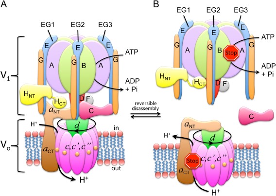
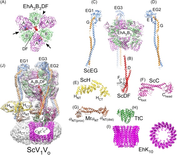
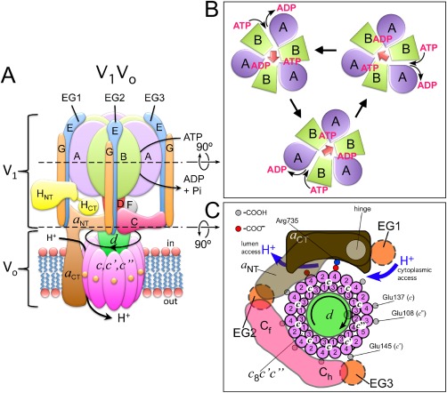
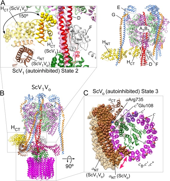
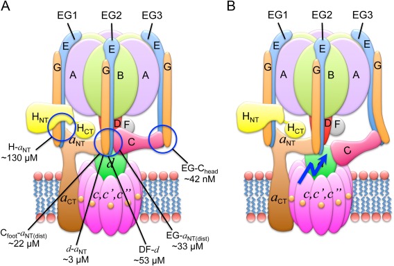
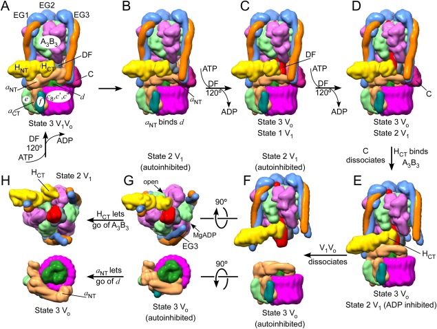
The vacuolar ATPase (V-ATPase; V1 Vo -ATPase) is a large multisubunit proton pump found in the endomembrane system of all eukaryotic cells where it acidifies the lumen of subcellular organelles including lysosomes, endosomes, the Golgi apparatus, and clathrin-coated vesicles. V-ATPase function is essential for pH and ion homeostasis, protein trafficking, endocytosis, mechanistic target of rapamycin (mTOR), and Notch signaling, as well as hormone secretion and neurotransmitter release. V-ATPase can also be found in the plasma membrane of polarized animal cells where its proton pumping function is involved in bone remodeling, urine acidification, and sperm maturation. Aberrant (hypo or hyper) activity has been associated with numerous human diseases and the V-ATPase has therefore been recognized as a potential drug target. Recent progress with moderate to high-resolution structure determination by cryo electron microscopy and X-ray crystallography together with sophisticated single-molecule and biochemical experiments have provided a detailed picture of the structure and unique mode of regulation of the V-ATPase. This review summarizes the recent advances, focusing on the structural and biophysical aspects of the field.

Keywords: V-ATPase; V1Vo-ATPase; X-ray crystallography; cryo electron microscopy; protein structure; protein-protein interactions; reversible disassembly; rotary catalysis; rotary motor enzyme; vacuolar ATPase.


Figures

Figure 1
Figure 1
V‐ATPase subunit arrangement and regulation by reversible dissociation. (A) Subunit architecture of yeast V‐ATPase. (B) Regulation by reversible dissociation. In yeast, under conditions of starvation, V1‐ATPase disengages from the membrane bound Vo and the activity of both sectors is silenced. Activity silencing is accompanied by a large conformational change in V1's H subunit (HCT) and the N‐terminal domain of the Vo a subunit (a NT). Illustration adapted from Ref. 73.
Figure 2
Figure 2
Overall subunit architecture of holo V‐ATPase. (A) Crystal structure of EhA3B3DF (3vr6; a side‐view of the structure is also shown on the right).47 The arrows indicate closed catalytic sites with AMPPNP bound and the arrowhead points to the open site. (B) Crystal structure of ScDF (4rnd).48 (C,D) Crystal structures of ScEGChead in two conformations (4dl0, 4efa; Chead not shown).49 (E) Crystal structure of ScH (1ho8).50 (F) Crystal structure of ScC (1u7l).51 (G) Crystal structure of the a NT homolog from M. ruber (Mra; 3rrk).52 (H) Crystal structure of the d homolog from T. thermophilus (TtC; 1r5z).103 (I) Crystal structure of the proteolipid ring from E. hirae (EhK10; 2bl2)53. (J) CryoEM map of ScV1Vo (emd‐6284)45 with fitted coordinate models of individual subunits and subcomplexes (3j9t).45 The overall model was generated from individual crystal structures of yeast subunits and homology models generated by threading yeast primary sequences into crystal structures of bacterial A‐ATPase subunits.
Figure 3
Figure 3
V‐ATPase mechanism. (A) V‐ATPase consists of two motors, a V1‐ATPase and a Vo proton channel. (B) ATP hydrolysis at three of the six AB interfaces drives counterclockwise rotation of the central DF rotor (A3B3 seen towards the cytosol). (C) Rotation of the central DF rotor drives rotation of the proteolipid ring past the essential arginine residue in a CT. Protons enter a cytoplasmic half‐channel and, after being carried 360º on lipid exposed glutamate residues on the c subunits, are released into the lumenal half‐channel.
Figure 4
Figure 4
V‐ATPase conformational changes accompanying reversible disassembly. (A) Conformational changes in the auto‐inhibited ScV1‐ATPase (5d80).71 Upon holo enzyme dissociation, the C‐terminus of subunit H (HCT) is released from its binding site on a NT (in V1Vo) and rotates 150º to bind at the bottom of the A3B3 hexamer (in V1). HCT binds to the B subunit of the open catalytic site, thereby stabilizing inhibitory ADP in another site.71 The inhibitory loop is in spacefill and the α helix that binds a NT in V1Vo is in orange. (B) CryoEM model of holo ScV1Vo 45 (3j9u). (C) Conformational changes in the Vo proton channel (5tj5)26. Upon release of V1‐ATPase from the membrane, the N‐terminus of subunit a (a NT) moves from its ternary interface with Cfoot and EG2 to form a new binding interface with subunit d 26, 73, 88 and the c‐ring is positioned so that Glu108 of isoform c″ is in contact with the essential Arg735 in a CT.
Figure 5
Figure 5
Interactions at the V1–Vo interface. (A) The affinities of the individual interactions have been measured using isothermal titration calorimetry of recombinant yeast V‐ATPase subunits.48, 96, 97 (B) Spring‐loading of EG3. It has been suggested that the tension in EG3 would prevent re‐forming of the EG2–a NT(distal)–Cfoot ternary interface after one of these interactions is broken to initiate disassembly.49
Figure 6
Figure 6
Model of the structural changes along the steps of holo V‐ATPase disassembly. (A) It has been proposed that disassembly is initiated in state 3 V1Vo. 26 (B) The ternary a NT–EG2–Cfoot interaction is destabilized, allowing a NT to bind subunit d. (C,D) Two uncoupled catalytic steps convert V1 into the state 2 conformation. (E) Subunit C dissociates and HCT rotates 150º to bind at the open catalytic site. V1 is now in state 2 and autoinhibited and Vo is in state 3, also autoinhibited. (F) V1Vo dissociates. (G) Views towards to bottom of autoinhibited V1, illustrating HCT's binding site on A3B3DF, and towards the top of Vo, showing the interaction of a NT with subunit d. (H) Reassembly must involve release of HCT and a NT from their autoinhibitory positions and one catalytic turnover of V1 to return both sectors into matching state 3 conformations. Note that the starting conformation of V1Vo and the order of the steps are not firmly established. See also Supporting Information Fig. S1 for an animation of the process.

Similar articles

Cited by

References

    1. Forgac M (2007) Vacuolar ATPases: rotary proton pumps in physiology and pathophysiology. Nat Rev Mol Cell Biol 8:917–929. - PubMed
    1. Kane PM (2006) The where, when, and how of organelle acidification by the yeast vacuolar H+‐ATPase. Microbiol Mol Biol Rev 70:177–191. - PMC - PubMed
    1. Marshansky V, Futai M (2008) The V‐type H+‐ATPase in vesicular trafficking: targeting, regulation and function. Curr Opin Cell Biol 20:415–426. - PMC - PubMed
    1. Graham LA, Flannery AR, Stevens TH (2003) Structure and assembly of the yeast V‐ATPase. J Bioenerg Biomembr 35:301–312. - PubMed
    1. Zoncu R, Bar‐Peled L, Efeyan A, Wang S, Sancak Y, Sabatini DM (2011) mTORC1 senses lysosomal amino acids through an inside‐out mechanism that requires the vacuolar H(+)‐ATPase. Science 334:678–683. - PMC - PubMed

MeSH terms

Substances

LinkOut - more resources