. 2016 Nov 20;34(33):4030-4039.
doi: 10.1200/JCO.2016.67.1222. Epub 2016 Oct 31.

Prospective Longitudinal Analysis of 2-Hydroxyglutarate Magnetic Resonance Spectroscopy Identifies Broad Clinical Utility for the Management of Patients With IDH-Mutant Glioma

Affiliations

Prospective Longitudinal Analysis of 2-Hydroxyglutarate Magnetic Resonance Spectroscopy Identifies Broad Clinical Utility for the Management of Patients With IDH-Mutant Glioma

Changho Choi et al. J Clin Oncol. .

Abstract

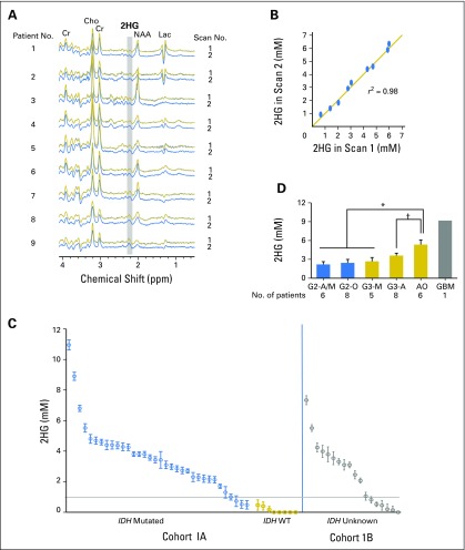
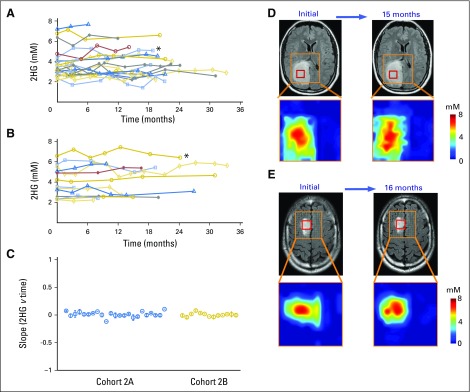
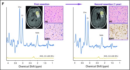
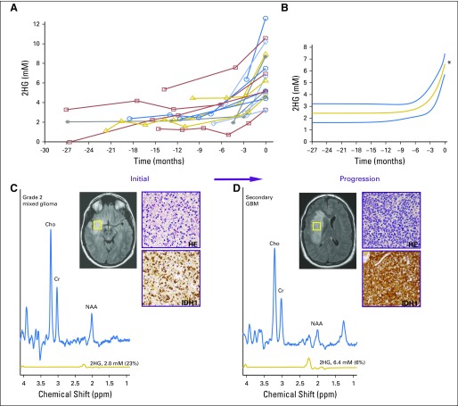
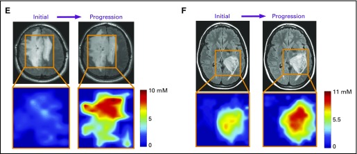
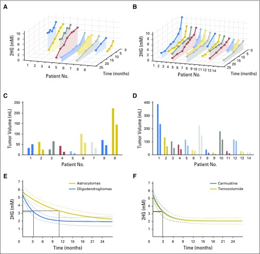
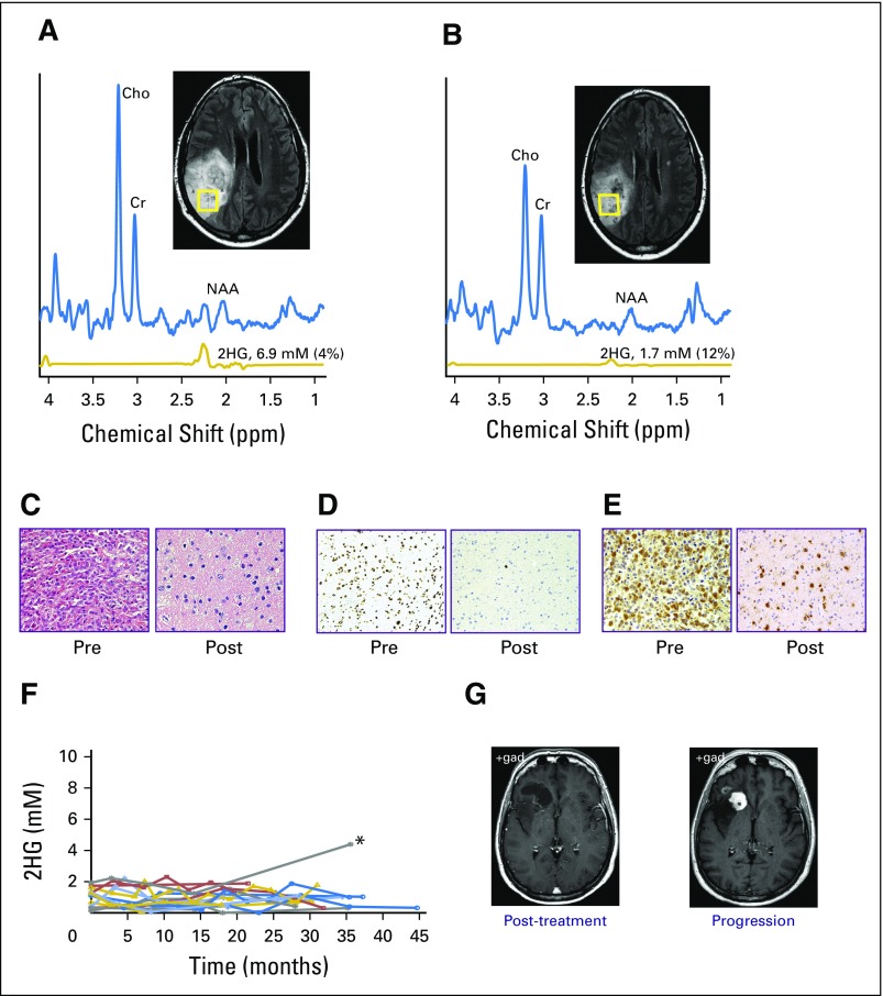
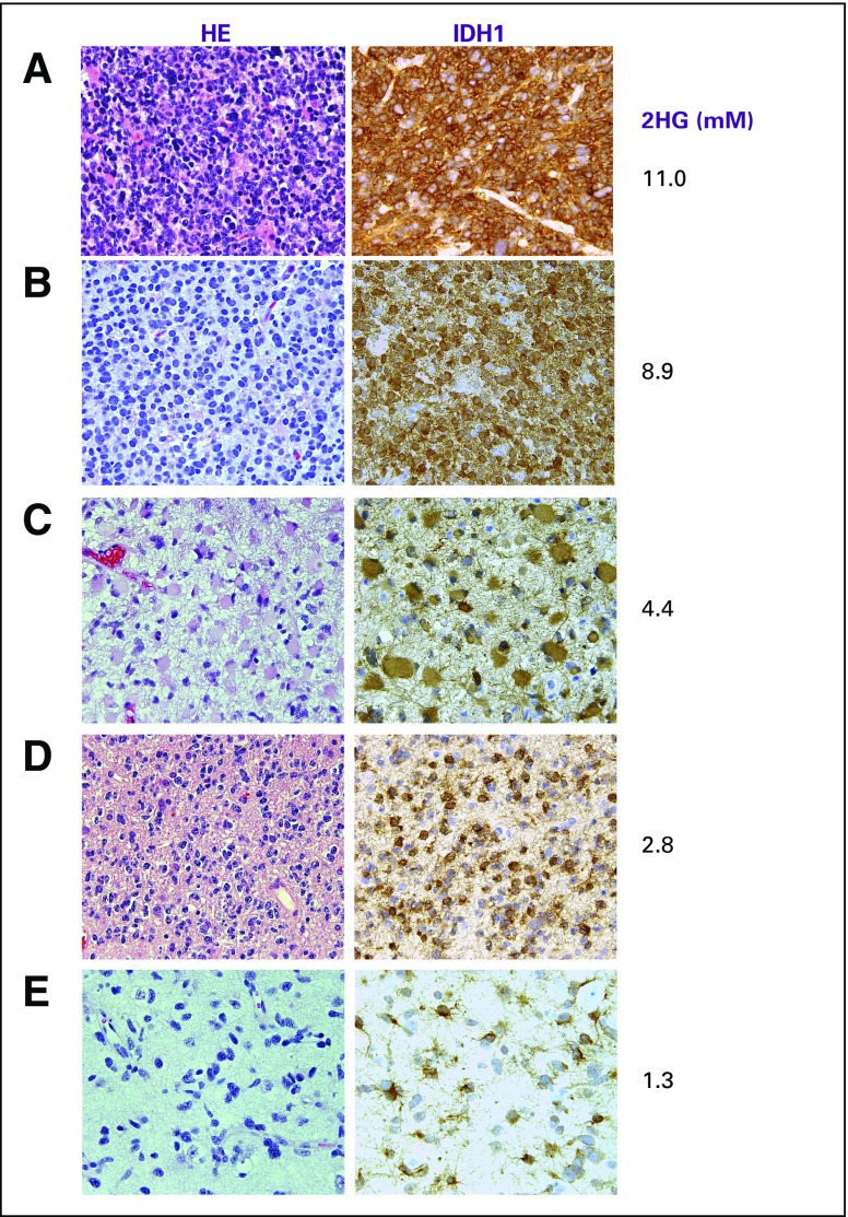
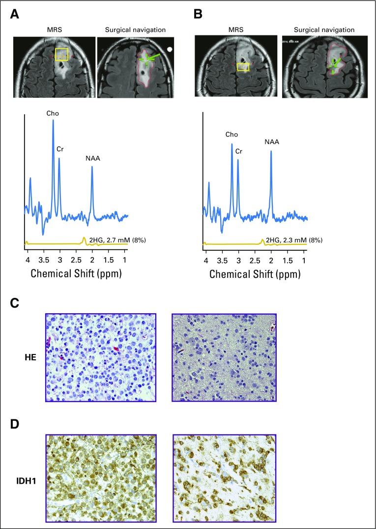
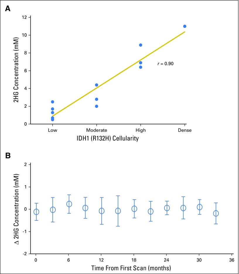
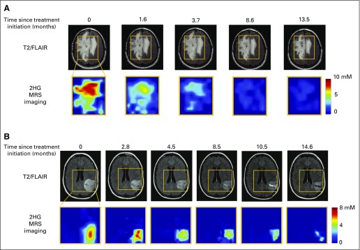
Purpose Proton magnetic resonance spectroscopy (MRS) of the brain can detect 2-hydroxyglutarate (2HG), the oncometabolite produced in neoplasms harboring a mutation in the gene coding for isocitrate dehydrogenase ( IDH). We conducted a prospective longitudinal imaging study to determine whether quantitative assessment of 2HG by MRS could serve as a noninvasive clinical imaging biomarker for IDH-mutated gliomas. Patients and Methods 2HG MRS was performed in 136 patients using point-resolved spectroscopy at 3 T in parallel with standard clinical magnetic resonance imaging and assessment. Data were analyzed in patient cohorts representing the major phases of the glioma clinical course and were further subgrouped by histology and treatment type to evaluate 2HG. Histologic correlations were performed. Results Quantitative 2HG MRS was technically and biologically reproducible. 2HG concentration > 1 mM could be reliably detected with high confidence. During the period of indolent disease, 2HG concentration varied by less than ± 1 mM, and it increased sharply with tumor progression. 2HG concentration was positively correlated with tumor cellularity and significantly differed between high- and lower-grade gliomas. In response to cytotoxic therapy, 2HG concentration decreased rapidly in 1p/19q codeleted oligodendrogliomas and with a slower time course in astrocytomas and mixed gliomas. The magnitude and time course of the decrease in 2HG concentration and magnitude of the decrease in tumor volume did not differ between oligodendrogliomas treated with temozolomide or carmustine. Criteria for 2HG MRS were established to make a presumptive molecular diagnosis of an IDH mutation in gliomas technically unable to undergo a surgical procedure. Conclusion 2HG concentration as measured by MRS was reproducible and reliably reflected the disease state. These data provide a basis for incorporating 2HG MRS into clinical management of IDH-mutated gliomas.


Conflict of interest statement

Authors’ disclosures of potential conflicts of interest are found in the article online at www.jco.org. Author contributions are found at the end of this article.

Figures

Fig 1.
Fig 1.
Baseline characteristics of 2-hydroxyglutarate (2HG) as measured by magnetic resonance spectroscopy in patients with untreated tumors. (A) Test–retest analysis in nine patients, arranged in ascending order of 2HG concentration. In vivo spectra are shown for the first (scan 1) and second scans (scan 2). (B) Linear regression curve of 2HG concentration in scan 1 v 2. (C) 2HG concentration (± standard deviation [SD]) from 60 individual tumors in two patient groups; cohort 1A: those with a tissue diagnosis (n = 42), and cohort 1B: those without a tissue diagnosis (n = 18), shown from high to low 2HG concentration. The SD of each 2HG concentration estimate was obtained from the Cramer–Rao lower bound value in the spectral fitting. (D) Comparison of 2HG concentration among tumors grouped by grade and histologic diagnosis. A, astrocytoma; AO, anaplastic oligodendroglioma (includes patients with progressive grade 2 oligodendroglioma who had new enhancement and increased tumor size on T2/FLAIR sequence); Cho, choline; Cr, creatine; G, grade; GBM, glioblastoma; Lac, lactate; M, mixed glioma; NAA, N-acetyl aspartate; O, oligodendroglioma; T2/FLAIR, T2-weighted fluid-attenuated inversion recovery; WT, wild type. (*) P < .001. (†) P = .02.
Fig 2.
Fig 2.
Stable 2-hydroxyglutarate (2HG) concentration in patients with nonenhancing indolent disease. (A, B) Single voxel MRS scans were analyzed serially in individual patients in the (A) IDH-mutated group (cohort 2A; n = 23) and (B) no tissue diagnosis group (cohort 2B; n = 13). Each curve represents one patient, and each data point was obtained from a separate magnetic resonance (MR) scan with spectroscopy (MRS) and 2HG concentration determination. Month 0 was the first scan at study enrollment. (C) Slope (± standard deviation) of 2HG concentration versus month for each patient curve from (A) and (B). (D, E) Multivoxel imaging spectra were obtained from patients denoted by (*) at two separate time points in (A) and (B). Interval between scans was 15 and 16 months, respectively. T2/FLAIR images are overlaid with the multivoxel grid (5 × 5 mm2, with 15-mm slice thickness). Red square shows the location of the single voxel used to generate the data for the patients in (A) and (B). The 2HG concentration color maps are shown for each grid (range, 0 to 8 mM). (F) Comparison of preoperative MRS (voxel size, 20 × 20 × 20 mm3), 2HG concentration, postoperative hematoxylin and eosin (HE), and isocitrate dehydrogenase 1 (IDH1)/R132H immunoreactivity at two time points for a patient with grade 2 astrocytoma. Numbers in parentheses indicate the Cramer–Rao lower bounds of the 2HG measurement. Changes in any of the parameters were negligible 1 year after initial evaluation. Histologic images are ×200 magnification. Cho, choline; Cr, creatine; NAA, N-acetyl aspartate; T2/FLAIR, T2-weighted fluid-attenuated inversion recovery.
Fig 2.
Fig 2.
Stable 2-hydroxyglutarate (2HG) concentration in patients with nonenhancing indolent disease. (A, B) Single voxel MRS scans were analyzed serially in individual patients in the (A) IDH-mutated group (cohort 2A; n = 23) and (B) no tissue diagnosis group (cohort 2B; n = 13). Each curve represents one patient, and each data point was obtained from a separate magnetic resonance (MR) scan with spectroscopy (MRS) and 2HG concentration determination. Month 0 was the first scan at study enrollment. (C) Slope (± standard deviation) of 2HG concentration versus month for each patient curve from (A) and (B). (D, E) Multivoxel imaging spectra were obtained from patients denoted by (*) at two separate time points in (A) and (B). Interval between scans was 15 and 16 months, respectively. T2/FLAIR images are overlaid with the multivoxel grid (5 × 5 mm2, with 15-mm slice thickness). Red square shows the location of the single voxel used to generate the data for the patients in (A) and (B). The 2HG concentration color maps are shown for each grid (range, 0 to 8 mM). (F) Comparison of preoperative MRS (voxel size, 20 × 20 × 20 mm3), 2HG concentration, postoperative hematoxylin and eosin (HE), and isocitrate dehydrogenase 1 (IDH1)/R132H immunoreactivity at two time points for a patient with grade 2 astrocytoma. Numbers in parentheses indicate the Cramer–Rao lower bounds of the 2HG measurement. Changes in any of the parameters were negligible 1 year after initial evaluation. Histologic images are ×200 magnification. Cho, choline; Cr, creatine; NAA, N-acetyl aspartate; T2/FLAIR, T2-weighted fluid-attenuated inversion recovery.
Fig 3.
Fig 3.
Tumor progression is associated with an increase in 2-hydroxyglutarate (2HG) concentration. (A) Eighteen patients had at least one baseline scan with 2HG concentration measurement before Response Assessment in Neuro-Oncology–confirmed tumor progression, at the 0-months time point. Data prior to progression (0-months time point) are represented by negative months. Each curve represents one patient, and each data point was obtained from a separate magnetic resonance (MR) scan with spectroscopy (MRS) and 2HG concentration determination. (B) Curve fitting with an exponential function. The fit (2.42 + 4.17e−0.41 month) is shown together with 95% CIs (blue lines). The coefficient of determination (r2) was 0.49. The mean 2HG concentration at progression (6.59 mM) was significantly greater than the mean baseline 2HG concentration (2.42 mM). (C) A representative patient from (A) with grade 2 mixed glioma is shown at the time of initial scanning after biopsy (MRS voxel size, 20 × 20 × 20 mm2). Hematoxylin and eosin (HE) staining shows moderate cellularity, and isocitrate dehydrogenase 1 (IDH1)/R132H shows strong immunoreactivity. (D) Same patient in (C) at time of progression (preoperative MRS voxel size, 20 × 20 × 20 mm3; 2HG concentration, 6.4 mM). Diagnosis at second surgery was glioblastoma. HE staining shows high cellularity, with strong IDH1/R132H immunoreactivity. Numbers in parentheses indicate the 2HG Cramer–Rao lower bounds of the 2HG measurements. (E, F) Multivoxel imaging of two patients from (A) at two time points: initial scan and progression (time 0). Outline of grid overlays T2/FLAIR images, and 2HG concentration color map is shown for each time point. (E) Untreated grade 2 oligodendroglioma (1p/19q codeleted, IDH2 R172K mutated), which progressed during surveillance. Minimal gadolinium enhancement in the right frontal lobe on clinical MR imaging (MRI; not shown). Time between initial and progression scan was 5 months. 2HG concentration color map range is 0 to 10 mM. (F) Recurrent grade 3 astrocytoma (IDH1 R132H mutated), which progressed during temozolomide treatment. Time between initial and progression scans was 4 months. 2HG concentration color map range is 0 to 11 mM. Clinical scan showed a 5-mm focus of gadolinium enhancement on clinical MRI at progression (not shown). Histologic images are ×200 magnification. Cho, choline; Cr, creatine; NAA, N-acetyl aspartate; T2/FLAIR, T2-weighted fluid-attenuated inversion recovery. (*) P < .001. (Continued)
Fig 3.
Fig 3.
Tumor progression is associated with an increase in 2-hydroxyglutarate (2HG) concentration. (A) Eighteen patients had at least one baseline scan with 2HG concentration measurement before Response Assessment in Neuro-Oncology–confirmed tumor progression, at the 0-months time point. Data prior to progression (0-months time point) are represented by negative months. Each curve represents one patient, and each data point was obtained from a separate magnetic resonance (MR) scan with spectroscopy (MRS) and 2HG concentration determination. (B) Curve fitting with an exponential function. The fit (2.42 + 4.17e−0.41 month) is shown together with 95% CIs (blue lines). The coefficient of determination (r2) was 0.49. The mean 2HG concentration at progression (6.59 mM) was significantly greater than the mean baseline 2HG concentration (2.42 mM). (C) A representative patient from (A) with grade 2 mixed glioma is shown at the time of initial scanning after biopsy (MRS voxel size, 20 × 20 × 20 mm2). Hematoxylin and eosin (HE) staining shows moderate cellularity, and isocitrate dehydrogenase 1 (IDH1)/R132H shows strong immunoreactivity. (D) Same patient in (C) at time of progression (preoperative MRS voxel size, 20 × 20 × 20 mm3; 2HG concentration, 6.4 mM). Diagnosis at second surgery was glioblastoma. HE staining shows high cellularity, with strong IDH1/R132H immunoreactivity. Numbers in parentheses indicate the 2HG Cramer–Rao lower bounds of the 2HG measurements. (E, F) Multivoxel imaging of two patients from (A) at two time points: initial scan and progression (time 0). Outline of grid overlays T2/FLAIR images, and 2HG concentration color map is shown for each time point. (E) Untreated grade 2 oligodendroglioma (1p/19q codeleted, IDH2 R172K mutated), which progressed during surveillance. Minimal gadolinium enhancement in the right frontal lobe on clinical MR imaging (MRI; not shown). Time between initial and progression scan was 5 months. 2HG concentration color map range is 0 to 10 mM. (F) Recurrent grade 3 astrocytoma (IDH1 R132H mutated), which progressed during temozolomide treatment. Time between initial and progression scans was 4 months. 2HG concentration color map range is 0 to 11 mM. Clinical scan showed a 5-mm focus of gadolinium enhancement on clinical MRI at progression (not shown). Histologic images are ×200 magnification. Cho, choline; Cr, creatine; NAA, N-acetyl aspartate; T2/FLAIR, T2-weighted fluid-attenuated inversion recovery. (*) P < .001. (Continued)
Fig 4.
Fig 4.
Decrease in 2-hydroxyglutarate (2HG) concentration in response to treatment. Twenty-three patients were observed during treatment for a median of 15 months (range, 6 to 24 months), with a median of five scans (range, two to 10 scans) per patient. (A, B) Data are presented for each patient over time in two groups: (A) astrocytomas and mixed gliomas and (B) oligodendrogliomas. The first point of each curve is the immediate pretreatment 2HG concentration. Patients are arranged from highest pretreatment 2HG concentration to lowest. Each point on the curve is the 2HG concentration (mM; y-axis) at a specific time point (months; z-axis). (C, D) Corresponding tumor volume (mL) for each patient in (A) and (B), with corresponding patient Nos. and colors. For each patient, there are two measurements that correspond to the time point of the first and last 2HG measurements on the curves in (A) and (B). (E) The fits from a mixed nonlinear regression analysis of the 2HG concentration data in (A) and (B), which are 1.9 + 3.7e−month/11 and 1.9 + 3.4e−month/3.3, respectively, are shown. The coefficients of determination (r2) are 0.58 and 0.46, respectively. In each case, the month marked by a black solid line indicates the time point (ie, decaying time constant) at which the exponent equals −1. There was a significant difference in the rate of decrease in 2HG concentration (oligodendrogliomas, 3.3 months v astrocytomas and mixed gliomas, 11.0 months; P = .01). (F) A mixed nonlinear regression analysis of the oligodendroglioma data was performed. Patients were subgrouped by chemotherapy regimen: three to six cycles of carmustine (n = 7) or 12 cycles of temozolomide (n = 7). The fits (2.0 + 3.5e−month/3.0 and 2.0 + 3.3e−month/3.0, respectively) are shown together with 95% CIs (dashed lines). For each group, the month marked by a black solid line indicates the time point (ie, decaying time constant) at which the exponent equals −1. There was no significant difference in the rate of decrease in 2HG concentration (P = .99).
Fig 5.
Fig 5.
(A to E) Pre- and post-treatment comparison of 2-hydroxyglutarate (2HG) concentration and histologic markers in a patient with grade 3 oligodendroglioma (1p/19q codeleted, IDH1 R132H mutated). (A) Single-voxel magnetic resonance spectroscopy (MRS) data (voxel size, 20 × 20 × 20 mm3) with 2HG concentration measurement were obtained before initiation of carmustine (6.9 mM) and (B) 1 year after completing treatment (1.7 mM). Surgery was performed after the scan in (B). Numbers in parentheses indicate the 2HG Cramer–Rao lower bounds. (C) Hematoxylin and eosin staining shows high cellularity in the pretreatment biopsy, with marked decrease in the resected tumor post-treatment, (D) corresponding to a decrease in the MIB-1 proliferation index from 30% (pretreatment) to 1% (post-treatment). (E) All tumor cells were strongly isocitrate dehydrogenase 1 R132H immunoreactive. (F) Individual scans from 15 patients who had completed all treatment before their first 2HG MRS scan at 0 months. 2HG concentration was ≤ 2 mM over the course of follow-up, (*) except in one patient, who experienced progression at 35 months (2HG concentration, 4.4 mM). The prior 2HG MRS scan was performed at month 14 (2HG concentration, 1.2 mM). (G) Representative corresponding clinical postgadolinium axial images are shown for the post-treatment scan performed 1 year before progression and at the time of progression, showing new gadolinium enhancement. Histologic images in (C) and (E) are ×200 magnification, and in (D) are ×100 magnification. Cho, choline; Cr, creatine; NAA, N-acetyl aspartate.
Fig A1.
Fig A1.
Comparison of cellularity, isocitrate dehydrogenase 1 (IDH1; R132H) immunoreactivity, and 2-hydroxyglutarate (2HG) concentration (mM) among various grades of glioma. Cellularity was assessed from hematoxylin and eosin (HE) staining. All images are ×200 magnification. (A) Glioblastoma (secondary) shows multiple mitoses with dense cellularity and strong IDH1/R132H immunoreactivity. (B) Grade 3 oligodendroglioma (1p/19q codeleted) shows high cellularity and strong IDH1/R132H immunoreactivity. (C) Grade 3 astrocytoma and (D) grade 2 oligodendroglioma have moderate cellularity, whereas IDH1/R132H staining intensity was unchanged. (E) Grade 2 astrocytoma demonstrated low cellularity with equivalent IDH1/R132H staining intensity. 2HG concentration is correlated with cellularity.
Fig A2.
Fig A2.
Tumor heterogeneity and histologic variations contribute to 2-hydroxyglutarate (2HG) level. Representative case of grade 2 oligodendroglioma (isocitrate dehydrogenase 1 [IDH1], R132H). Coronal T2/FLAIR images show locations of (A) anterior voxel (18 × 20 × 15 mm3) and (B) posterior voxel (13 × 18 × 15 mm3). For each location, the voxel placement for magnetic resonance (MR) spectroscopy (MRS; yellow box) is shown on the left image, and the location of surgical sampling (green hatch) is on the right image. MR spectra and 2HG concentration from the two voxels were similar. Numbers in parentheses indicate the 2HG Cramer–Rao lower bounds. (C) Hematoxylin and eosin (HE) staining shows nodule of high cellularity on the left, contrasted with HE showing a region of low cellularity on the right. (D) IDH1/R132H immunohistochemistry corresponding to the sections in (C). Regions of both histologic findings were found in the anterior and posterior voxels. 2HG concentration in each voxel represents the composite contribution of the cellularity within the region of tumor. Cho, choline; Cr, creatine; NAA, N-acetyl aspartate; T2/FLAIR, T2-weighted fluid-attenuated inversion recovery.
Fig A3.
Fig A3.
(A) Correlation of 2-hydroxyglutarate (2HG) concentration with tumor cellularity. Using the isocitrate dehydrogenase 1 (IDH1)/R132H–specific antibody, tumor cellularity was determined for each patient who had this specific mutation and underwent a surgical procedure within 1 month of the 2HG magnetic resonance spectroscopy (MRS) evaluation (Spearman rank correlation r = 0.90). (B) Stable 2HG concentration during period of tumor stability. Single-voxel MRS scans were analyzed serially in individual patients (shown in Figs 2A and 2B), and 2HG concentration was plotted over time. Month 0 was the first scan at enrollment in the study. Here we show the change in 2HG concentration between scans for the 36 patients. Means (± standard deviations) were calculated for consecutive 3-month periods. There were no statistical differences at any time point.
Fig A4.
Fig A4.
Mapping of the decrease in 2-hydroxyglutarate (2HG) concentration after treatment. A representative series of multivoxel images is shown for two patients after treatment. (A) Same patient shown in Fig 3E, who was treated with six cycles of carmustine at the time of progression (multivoxel grid dimension, 100 × 80 mm2). Grid removed to enable visualization of T2-weighted fluid-attenuated inversion recovery (T2/FLAIR) changes over time. (B) Patient with grade 3 astrocytoma treated with concurrent temozolomide and radiotherapy followed by 12 cycles of temozolomide at the time of progression (multivoxel grid dimension, 90 × 100 mm2). MRS, magnetic resonance spectroscopy.

References

    1. Parsons DW, Jones S, Zhang X, et al. An integrated genomic analysis of human glioblastoma multiforme. Science. 2008;321:1807–1812. - PMC - PubMed
    1. Yan H, Parsons DW, Jin G, et al. IDH1 and IDH2 mutations in gliomas. N Engl J Med. 2009;360:765–773. - PMC - PubMed
    1. Dang L, White DW, Gross S, et al. Cancer-associated IDH1 mutations produce 2-hydroxyglutarate. Nature. 2009;462:739–744. - PMC - PubMed
    1. Brat DJ, Verhaak RG, Aldape KD, et al. Comprehensive, integrative genomic analysis of diffuse lower-grade gliomas. N Engl J Med. 2015;372:2481–2498. - PMC - PubMed
    1. Eckel-Passow JE, Lachance DH, Molinaro AM, et al. Glioma groups based on 1p/19q, IDH, and TERT promoter mutations in tumors. N Engl J Med. 2015;372:2499–2508. - PMC - PubMed

Publication types

MeSH terms