Review
. 2017 Mar 1;127(3):737-749.
doi: 10.1172/JCI84417. Epub 2017 Mar 1.

Chikungunya virus: epidemiology, replication, disease mechanisms, and prospective intervention strategies

Review

Chikungunya virus: epidemiology, replication, disease mechanisms, and prospective intervention strategies

Laurie A Silva et al. J Clin Invest. .

Abstract

Chikungunya virus (CHIKV), a reemerging arbovirus, causes a crippling musculoskeletal inflammatory disease in humans characterized by fever, polyarthralgia, myalgia, rash, and headache. CHIKV is transmitted by Aedes species of mosquitoes and is capable of an epidemic, urban transmission cycle with high rates of infection. Since 2004, CHIKV has spread to new areas, causing disease on a global scale, and the potential for CHIKV epidemics remains high. Although CHIKV has caused millions of cases of disease and significant economic burden in affected areas, no licensed vaccines or antiviral therapies are available. In this Review, we describe CHIKV epidemiology, replication cycle, pathogenesis and host immune responses, and prospects for effective vaccines and highlight important questions for future research.


Conflict of interest statement

Conflict of interest: The authors have declared that no conflict of interest exists.

Figures

Figure 1
Figure 1. Geographic distribution of endemic CHIKV and its primary vectors, Ae. aegypti and Ae. albopictus.
Countries in which autochthonous cases of CHIKV have been reported are specified with colored symbols representing the distinct viral genotypes detected during outbreaks in that country. West African strains are indicated by purple triangles; Asian strains are indicated by green circles; East/South/Central African (ESCA) strains are indicated with blue squares; strains of the Indian Ocean lineage, a subtype of the ESCA clade, are indicated with blue squares with a cross; and strains whose genotype has not been determined are indicated with gray diamonds. Symbols are shaded to differentiate transmission prior to (darker hue) or after (lighter hue) the reemergence of the virus in the Indian Ocean (ECSA strain in 2005) and Pacific Islands (Asian strain in 2010). Symbols indicate the countries in which natural transmission has occurred and are not meant to indicate precise locations of outbreaks. Overlayed with CHIKV distribution is the geographic range of the two primary vectors responsible for urban mosquito-human-mosquito transmission of the virus. Range of Ae. aegypti is indicated in red; range of Ae. albopictus is indicated in yellow; and areas where both mosquito species are present are indicated in orange. Endemic CHIKV data were obtained from numerous PubMed publications (, , , , , –, , , –62). Range of mosquito data was obtained from (63).
Figure 2
Figure 2. CHIKV replication cycle in mammalian cells.
(i) E2 binds to the cell surface via an unknown receptor and possibly glycosaminoglycans as attachment factors. (ii) CHIKV enters the cell through clathrin-mediated endocytosis. Acidification of endosomes leads to insertion of the fusion peptide in E1 into the endosomal membrane. (iii) Fusion of the viral envelope and endosomal membrane releases nucleocapsid into the cytosol. (iv) Disassembly of the nucleocapsid liberates positive-sense genomic RNA and nonstructural protein (nsP) translation occurs. (v) Four nsPs, together with genomic RNA and presumably host proteins, assemble at the plasma membrane (PM) and modify it to form viral replication compartments (spherules) containing viral dsRNA. nsP1–4 function as a replicase and localize to the spherule neck to generate genomic, antigenomic, and subgenomic vRNAs. (vi) Spherule internalization allows formation of large cytopathic vacuoles (CPV-1) that house multiple spherules. Spherules at the PM or within CPV-I are fully functional. (vii) Translation of subgenomic RNA produces the structural polyprotein, and capsid autoproteolysis releases free capsid into the cytoplasm. Translocation of E3-E2-6K-E1/E2-E2-TK polyproteins into the ER. E2/E1 are posttranslationally modified, transit the secretory system, and are deposited at the PM. (viii) Interaction of capsid and genomic RNA leads to formation of icosahedral nucleocapsids. (ix) Nucleocapsids assemble with E2/E1 at the PM, resulting in budding of mature progeny virions. (x) Later in infection, CPV-IIs form, containing hexagonal lattices of E2/E1 and are studded with nucleocapsids. (xi) CPV-IIs likely serve as transport vehicles and assembly sites for structural proteins, allowing formation of mature virions and egress.
Figure 3
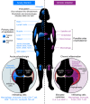
Figure 3. Model of acute and chronic CHIKV pathogenesis.
Acute CHIKV infection begins with transmission of the virus via a bite of an infected mosquito to the skin, where it replicates in susceptible cells, including fibroblasts and macrophages. The virus disseminates through lymphatics and bloodstream to typical (solid) and atypical (hatched) sites of primary replication (indicated in blue). Acute infection elicits an inflammatory response in infected tissues, characterized by an extensive infiltration of mainly macrophages and monocytes, but also neutrophils, NK cells, and lymphocytes in target tissues (indicated in blue), and by elaboration of a number of proinflammatory chemokines and cytokines. Within arthroskeletal tissues, synovial hyperplasia begins. Viral replication and host immune responses cause myalgia and polyarthralgia in distal joints. Chronic CHIKV disease can persist for months or years after acute infection but is often limited to more distal joints. Chronic disease is likely mediated by persistent virus and inflammation. Possible sites of CHIKV persistence include endothelial cells in the liver and other organs, mononuclear cells in the spleen, macrophages within the synovial fluid and surrounding tissues, and satellite cells within the muscle (indicated in purple). Within the chronically infected joint, the continued presence of a subset of infiltrating cells (mainly macrophages, monocytes, and lymphocytes) and specific proinflammatory mediators (IL-6, IL-8, and MCP-1) within the synovial fluid likely contribute to prolonged inflammatory disease. Chronic joint pathology resembles that in rheumatoid arthritis, with significant hyperplasia and angiogenesis. This model is based on human and animal studies.

Similar articles

Cited by

References

    1. Lumsden WH. An epidemic of virus disease in Southern Province, Tanganyika Territory, in 1952–53. II. General description and epidemiology. Trans R Soc Trop Med Hyg. 1955;49(1):33–57. doi: 10.1016/0035-9203(55)90081-X. - DOI - PubMed
    1. Robinson MC. An epidemic of virus disease in Southern Province, Tanganyika Territory, in 1952–53. I. Clinical features. Trans R Soc Trop Med Hyg. 1955;49(1):28–32. doi: 10.1016/0035-9203(55)90080-8. - DOI - PubMed
    1. Ross RW. The Newala epidemic. III. The virus: isolation, pathogenic properties and relationship to the epidemic. J Hyg (Lond) 1956;54(2):177–191. doi: 10.1017/S0022172400044442. - DOI - PMC - PubMed
    1. Thiberville SD, et al. Chikungunya fever: epidemiology, clinical syndrome, pathogenesis and therapy. Antiviral Res. 2013;99(3):345–370. doi: 10.1016/j.antiviral.2013.06.009. - DOI - PMC - PubMed
    1. Carey DE. Chikungunya and dengue: a case of mistaken identity? J Hist Med Allied Sci. 1971;26(3):243–262. - PubMed