

Comparative Skeletal Muscle Proteomics Using Two-Dimensional Gel Electrophoresis
- PMID: 28248237
- PMCID: PMC5217355
- DOI: 10.3390/proteomes4030027
Comparative Skeletal Muscle Proteomics Using Two-Dimensional Gel Electrophoresis
Abstract
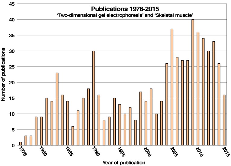
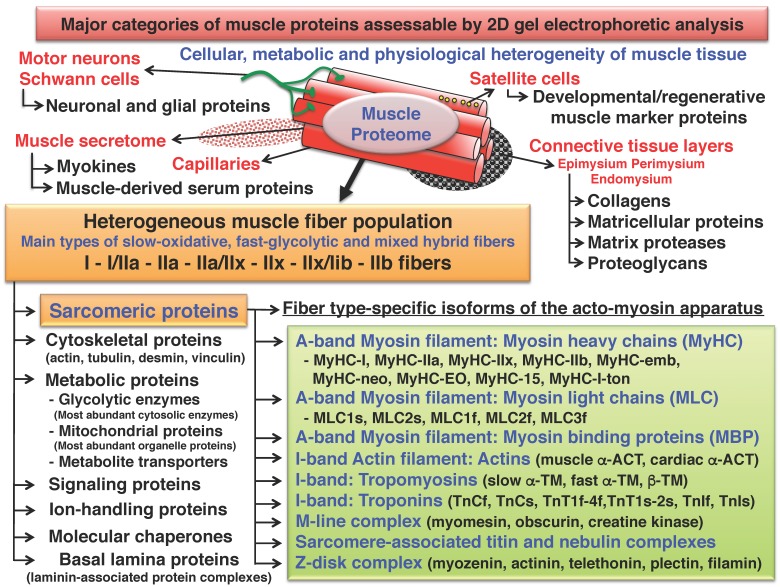
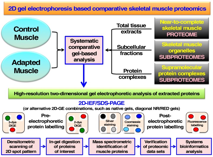
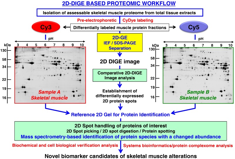
The pioneering work by Patrick H. O'Farrell established two-dimensional gel electrophoresis as one of the most important high-resolution protein separation techniques of modern biochemistry (Journal of Biological Chemistry1975, 250, 4007-4021). The application of two-dimensional gel electrophoresis has played a key role in the systematic identification and detailed characterization of the protein constituents of skeletal muscles. Protein changes during myogenesis, muscle maturation, fibre type specification, physiological muscle adaptations and natural muscle aging were studied in depth by the original O'Farrell method or slightly modified gel electrophoretic techniques. Over the last 40 years, the combined usage of isoelectric focusing in the first dimension and sodium dodecyl sulfate polyacrylamide slab gel electrophoresis in the second dimension has been successfully employed in several hundred published studies on gel-based skeletal muscle biochemistry. This review focuses on normal and physiologically challenged skeletal muscle tissues and outlines key findings from mass spectrometry-based muscle proteomics, which was instrumental in the identification of several thousand individual protein isoforms following gel electrophoretic separation. These muscle-associated protein species belong to the diverse group of regulatory and contractile proteins of the acto-myosin apparatus that forms the sarcomere, cytoskeletal proteins, metabolic enzymes and transporters, signaling proteins, ion-handling proteins, molecular chaperones and extracellular matrix proteins.
Keywords: difference in-gel electrophoresis; isoelectric focusing; mass spectrometry; muscle fiber type; muscle plasticity; muscle proteomics; muscular atrophy; polyacrylamide gel electrophoresis; protein separation; skeletal muscle.
Conflict of interest statement
The authors declare no conflict of interest.
Figures
Similar articles
-
Characterization of Contractile Proteins from Skeletal Muscle Using Gel-Based Top-Down Proteomics.Proteomes. 2019 Jun 20;7(2):25. doi: 10.3390/proteomes7020025. Proteomes. 2019. PMID: 31226838 Free PMC article. Review.
-
Two-dimensional polyacrylamide gel electrophoresis using flat-bed isoelectric focusing in the first dimension.Methods Mol Biol. 1988;3:217-31. doi: 10.1385/0-89603-126-8:217. Methods Mol Biol. 1988. PMID: 21400166
-
Comparative 3-Sample DIGE Analysis of Skeletal Muscles.Methods Mol Biol. 2018;1664:93-108. doi: 10.1007/978-1-4939-7268-5_9. Methods Mol Biol. 2018. PMID: 29019128
-
Comparative 3-Sample 2D-DIGE Analysis of Skeletal Muscles.Methods Mol Biol. 2023;2596:127-146. doi: 10.1007/978-1-0716-2831-7_11. Methods Mol Biol. 2023. PMID: 36378437
-
Current two-dimensional electrophoresis technology for proteomics.Proteomics. 2004 Dec;4(12):3665-85. doi: 10.1002/pmic.200401031. Proteomics. 2004. PMID: 15543535 Review.
Cited by
-
Molecular composition of skeletal muscle in infants and adults: a comparative proteomic and transcriptomic study.Sci Rep. 2024 Oct 3;14(1):22965. doi: 10.1038/s41598-024-74913-4. Sci Rep. 2024. PMID: 39362957 Free PMC article.
-
Fiber type diversity in skeletal muscle explored by mass spectrometry-based single fiber proteomics.Histol Histopathol. 2020 Mar;35(3):239-246. doi: 10.14670/HH-18-170. Epub 2019 Oct 15. Histol Histopathol. 2020. PMID: 31612964 Review.
-
Proteomic reference map for sarcopenia research: mass spectrometric identification of key muscle proteins of organelles, cellular signaling, bioenergetic metabolism and molecular chaperoning.Eur J Transl Myol. 2024 May 24;34(2):12565. doi: 10.4081/ejtm.2024.12565. Eur J Transl Myol. 2024. PMID: 38787292 Free PMC article.
-
Mass spectrometric identification of dystrophin, the protein product of the Duchenne muscular dystrophy gene, in distinct muscle surface membranes.Int J Mol Med. 2017 Oct;40(4):1078-1088. doi: 10.3892/ijmm.2017.3082. Epub 2017 Jul 27. Int J Mol Med. 2017. PMID: 28765879 Free PMC article.
-
Achyranthes bidentata extract protects chondrocytes functions through suppressing glycolysis and apoptosis via MAPK/AKT signaling axis.Am J Transl Res. 2020 Jan 15;12(1):142-152. eCollection 2020. Am J Transl Res. 2020. Retraction in: Am J Transl Res. 2025 May 15;17(5):4070. doi: 10.62347/VJKG9726. PMID: 32051743 Free PMC article. Retracted.
References
Publication types
LinkOut - more resources
Full Text Sources
Other Literature Sources
