

Reactive oxygen species generation by copper(II) oxide nanoparticles determined by DNA damage assays and EPR spectroscopy
- PMID: 28248593
- PMCID: PMC5494152
- DOI: 10.1080/17435390.2017.1293750
Reactive oxygen species generation by copper(II) oxide nanoparticles determined by DNA damage assays and EPR spectroscopy
Abstract
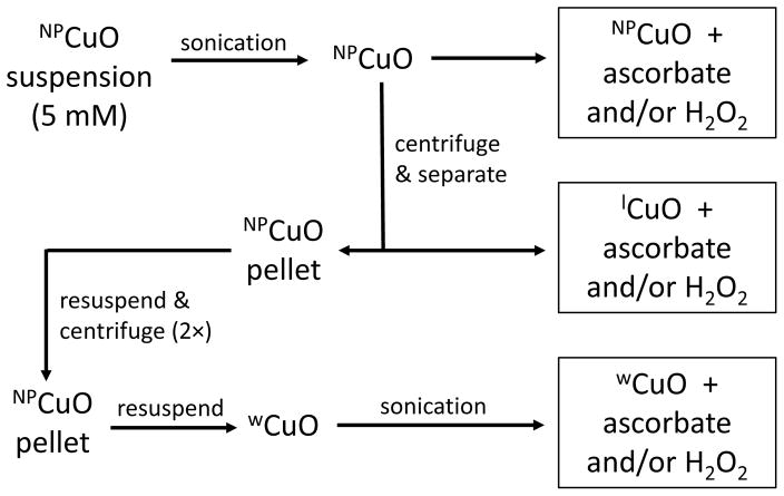
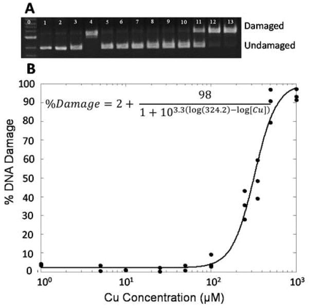
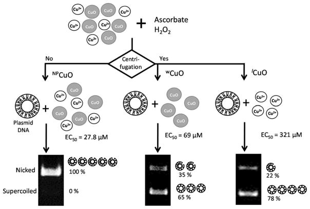
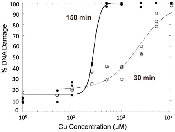
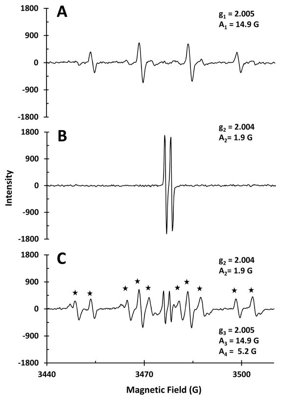
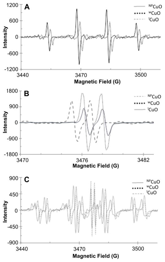
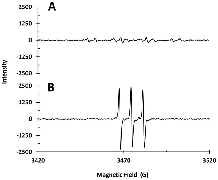
Copper(II) oxide nanoparticles (NPCuO) have many industrial applications, but are highly cytotoxic because they generate reactive oxygen species (ROS). It is unknown whether the damaging ROS are generated primarily from copper leached from the nanoparticles, or whether the nanoparticle surface plays a significant role. To address this question, we separated nanoparticles from the supernatant containing dissolved copper, and measured their ability to damage plasmid DNA with addition of hydrogen peroxide, ascorbate, or both. While DNA damage from the supernatant (measured using an electrophoresis assay) can be explained solely by dissolved copper ions, damage by the nanoparticles in the presence of ascorbate is an order of magnitude higher than can be explained by dissolved copper and must, therefore, depend primarily upon the nanoparticle surface. DNA damage is time-dependent, with shorter incubation times resulting in higher EC50 values. Hydroxyl radical (•OH) is the main ROS generated by NPCuO/hydrogen peroxide as determined by EPR measurements; NPCuO/hydrogen peroxide/ascorbate conditions generate ascorbyl, hydroxyl, and superoxide radicals. Thus, NPCuO generate ROS through several mechanisms, likely including Fenton-like and Haber-Weiss reactions from the surface or dissolved copper ions. The same radical species were observed when NPCuO suspensions were replaced with the supernatant containing leached copper, washed NPCuO, or dissolved copper solutions. Overall, NPCuO generate significantly more ROS and DNA damage in the presence of ascorbate than can be explained simply from dissolved copper, and the NPCuO surface must play a large role.
Keywords: DNA damage; Nanoparticles; nano-surfaces; nanotoxicology.
Conflict of interest statement
Figures
References
-
- Alves D, Santos CG, Paixao MW, Soares LC, De Souza D, Rodrigues OED, Braga AL. CuO nanoparticles: An efficient and recyclable catalyst for cross-coupling reactions of organic diselenides with aryl boronic acids. Tetrahedron Lett. 2009;50:6635–6638.
-
- Angelé-Martínez CGC, Brumaghim JL. Metal-mediated DNA damage and cell death: Mechanisms, detection methods, and cellular consequences. Metallomics. 2014;6:1358–1381. - PubMed
-
- Atha DH, Wang H, Petersen EJ, Cleveland D, Holbrook RD, Jaruga P, Dizdaroglu M, Xing B, Nelson BC. Copper oxide nanoparticle mediated DNA damage in terrestrial plant models. Environ Sci Technol. 2012;46:1819–1827. - PubMed
-
- Bartosz G. Use of spectroscopic probes for detection of reactive oxygen species. Clin Chim Acta. 2006;368:53–76. - PubMed
-
- Bielski BHJ. Reevaluation of the spectral and kinetic properties of HO2 and O2− free radicals. Photochem Photobiol. 1978;28:645–649.
MeSH terms
Substances
Grants and funding
LinkOut - more resources
Full Text Sources
Other Literature Sources
Miscellaneous