Case Reports
. 2017 Mar;96(9):e6225.
doi: 10.1097/MD.0000000000006225.

A case of mixed adenoneuroendocrine carcinoma of the pancreas: Immunohistochemical analysis for histogenesis

Affiliations
Case Reports

A case of mixed adenoneuroendocrine carcinoma of the pancreas: Immunohistochemical analysis for histogenesis

Masaru Murata et al. Medicine (Baltimore). 2017 Mar.

Abstract

Rationale: Tumors with multiple histological features, such as adenocarcinomas and neuroendocrine carcinomas, were included as a new category of neuroendocrine carcinomas in the 2010 World Health Organization classification. We recently experienced a rare case of a pancreatic carcinoma with both adenocarcinoma and neuroendocrine carcinoma components, a so-called mixed adenoneuroendocrine carcinoma.

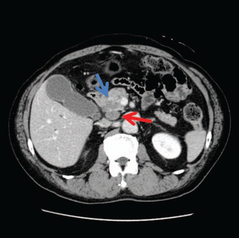
Patient concerns and diagnosis: A 66-year-old man was referred to our hospital with obstructive jaundice. Contrast-enhanced computed tomography images indicated a tumor located at the pancreatic head measuring 3.0 × 2.5 cm in diameter and invading the common bile duct. Cytological examination of the bile juice obtained by endoscopic retrograde cholangiopancreatography revealed adenocarcinoma cells. Pancreaticoduodenectomy was performed safely as radical resection.

Interventions: Microscopically, the resected tumor consisted of tightly intermingled adenocarcinoma and neuroendocrine carcinoma components. On the immunohistochemical examination, p53 was ubiquitously positive in both components, whereas chromogranin A, synaptophysin and neuron-specific enolase, neuroendocrine markers, were limited to the neuroendocrine carcinoma component.

Outcomes: Thus, such features of both adenocarcinoma and neuroendocrine carcinoma observed microscopically and immunohistochemically seemed to indicate a composite tumor.

Lessons: The findings of this case suggest that adenocarcinoma and neuroendocrine carcinoma may be derived from a single cancer stem cell.


Conflict of interest statement

The authors have no funding and conflicts of interest to disclose.

Figures

Figure 1
Figure 1
Contrast-enhanced computed tomography images showing a tumor in the pancreatic head measuring 3.0 × 2.5 cm in diameter and invading the common bile duct (indicated by a blue arrow). Lymphadenopathy was observed posterior to the pancreatic head (red arrow).
Figure 2
Figure 2
Microscopic and immunohistochemical appearance of the tumor. (A) Primary tumor (hematoxylin and eosin staining, original magnification ×20). The primary lesion was composed of a well-differentiated adenocarcinoma component and a poorly differentiated NEC component, each tightly intermingled. (B) Immunohistochemical staining of the primary lesion for CEA (original magnification ×20). CEA was limited to the well-differentiated adenocarcinoma component of the primary lesion. (C) LN no. 12 exclusively contained the well-differentiated adenocarcinoma component (original magnification ×20). (D) LN no. 13 exclusively contained the poorly differentiated NEC component (original magnification ×20). (E) Immunohistochemical staining of the primary lesion for chromogranin A (original magnification ×20). Chromogranin was limited to the poorly differentiated NEC component of the primary lesion. (F) Immunohistochemical staining of the primary lesion for synaptophysin (original magnification ×20). Synaptophysin was limited to the poorly differentiated NEC component of the primary lesion. (G) Immunohistochemical staining of the primary lesion for NSE (original magnification ×20). NSE was limited to the poorly differentiated NEC component of the primary lesion. (H) Immunohistochemical staining of the primary lesion for p53 (original magnification ×20). p53 was ubiquitously expressed in both the well-differentiated adenocarcinoma and poorly differentiated NEC components of the primary lesion. The well-differentiated adenocarcinoma component can be seen on the right sides and the poorly differentiated NEC component can be seen on the left (E, F, G, and H). CEA = carcinoembryonic antigen, LN = lymph node, NEC = neuroendocrine carcinoma, NSE = neuron-specific enolase.

References

    1. Bosman FT, Carneiro F, Theise ND. Nomenclature and classification of neuroendocrine neoplasms of digestive system. WHO Classification of Tumors of the Digestive System 4th ed.Lyon: IARC; 2010. 13–4.
    1. Vanacker L, Smeets D, Hoorens A, et al. Mixed adenoneuroendocrine carcinoma of the colon: molecular pathogenesis and treatment. Anticancer Res 2014;34:5517–22. - PubMed
    1. Liu X-J, Feng J-S, Xiang W-Y, et al. Clinicopathological features of an ascending colon mixed adenoneuroendocrine carcinoma with clinical serosal invasion. Int J Clin Exp Pathol 2014;7:6395–8. - PMC - PubMed
    1. Gurzu S, Kadar Z, Bara T, et al. Mixed adenoneuroendocrine carcinoma of gastrointestinal tract: report of two cases. World J Gastroenterol 2015;21:1329–33. - PMC - PubMed
    1. Zhang W, Xiao W, Ma H, et al. Neuroendocrine liver metastasis in gastric mixed adenoneuroendocrine carcinoma with trilineage cell differentiation: a case report. Int J Clin Exp Pathol 2014;7:6333–8. - PMC - PubMed

Publication types