. 2017 Mar 1;13(3):e1005406.
doi: 10.1371/journal.pcbi.1005406. eCollection 2017 Mar.

A magnesium-induced triplex pre-organizes the SAM-II riboswitch

Affiliations

A magnesium-induced triplex pre-organizes the SAM-II riboswitch

Susmita Roy et al. PLoS Comput Biol. .

Abstract

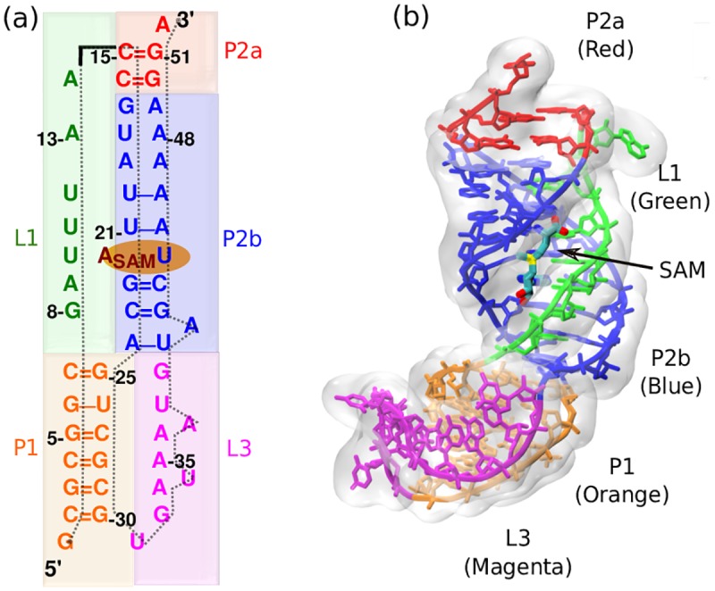
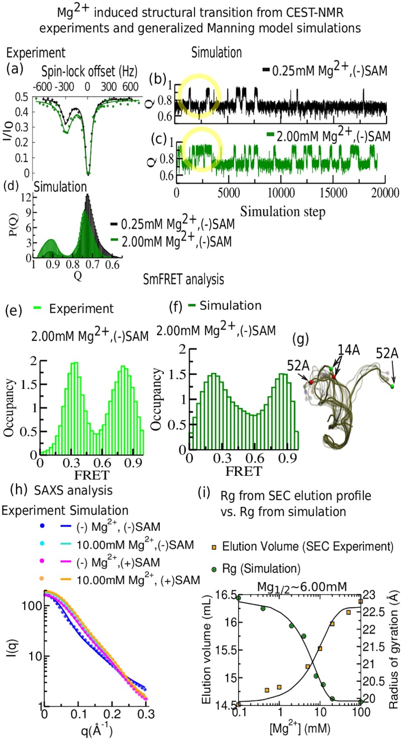
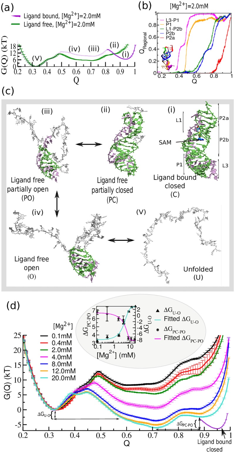
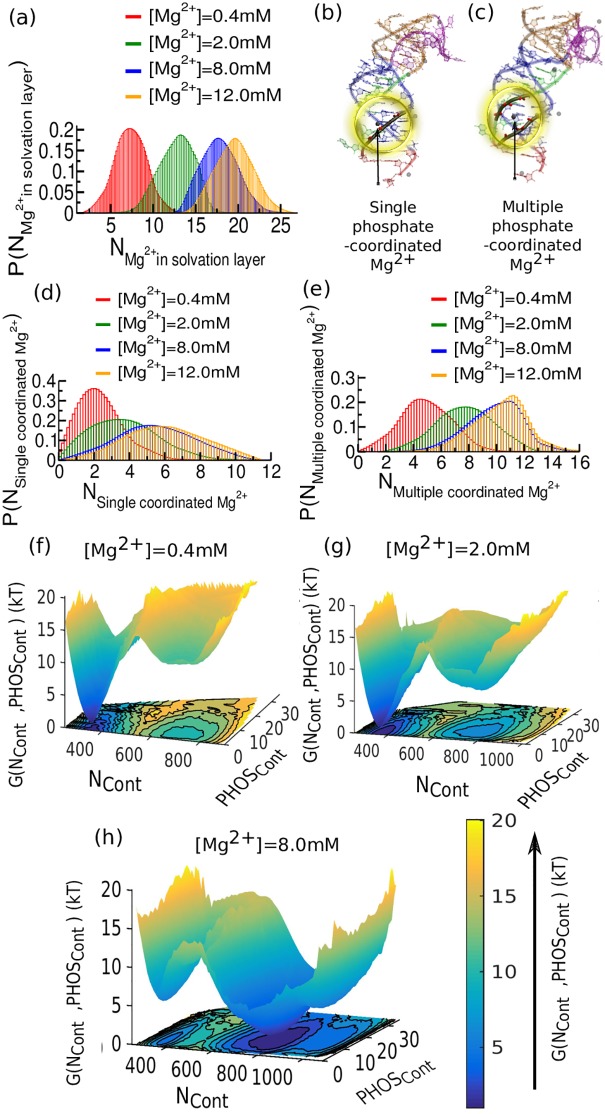
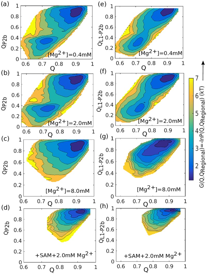
Our 13C- and 1H-chemical exchange saturation transfer (CEST) experiments previously revealed a dynamic exchange between partially closed and open conformations of the SAM-II riboswitch in the absence of ligand. Here, all-atom structure-based molecular simulations, with the electrostatic effects of Manning counter-ion condensation and explicit magnesium ions are employed to calculate the folding free energy landscape of the SAM-II riboswitch. We use this analysis to predict that magnesium ions remodel the landscape, shifting the equilibrium away from the extended, partially unfolded state towards a compact, pre-organized conformation that resembles the ligand-bound state. Our CEST and SAXS experiments, at different magnesium ion concentrations, quantitatively confirm our simulation results, demonstrating that magnesium ions induce collapse and pre-organization. Agreement between theory and experiment bolsters microscopic interpretation of our simulations, which shows that triplex formation between helix P2b and loop L1 is highly sensitive to magnesium and plays a key role in pre-organization. Pre-organization of the SAM-II riboswitch allows rapid detection of ligand with high selectivity, which is important for biological function.


Conflict of interest statement

The authors have declared that no competing interests exist.

Figures

Fig 1
Fig 1. Secondary and tertiary structure of SAM-II riboswitch in ligand-bound state (pdb:2QWY).
(a) Sequence-aligned secondary structure of the SAM-II riboswitch where base pair and stacking interactions are indicated. There are three helices and two strands highlighted with different colors. (P1: Orange, P2a: Red, P2b: Blue, L1: Green, L3: Magenta). (b) Tertiary structure displays triple helix between helix P2b and loop L1.
Fig 2
Fig 2. Detailed agreement between 13C CEST, smFRET, SAXS, and SEC elution profiles with the generalized Manning model corroborates pre-organization by Mg2+.
(a)-(d) Comparison between CEST-NMR experiments and Manning model simulations. (a) 13C CEST profiles for C43-C6 of SAM-II riboswitch in the ligand-free situation in the presence of two different concentrations of Mg2+ (black dots: 0.25 mM, green dots: 2.0 mM) at B1 field of 17.5 Hz. The population distribution involving two major states was obtained by fitting each 13C CEST profile (solid lines) into a two-state model. (b) The fraction of native contacts (Q) dynamics extracted from simulations indicate the transition between two major states at 0.25 mM. (c) Same as (b) for 2.0 mM Mg2+. (d) The fraction of native contact population histograms at these two concentrations. (e)-(f) Steady state smFRET analysis from fluorescently labeled experiments and theoretical prediction from simulations. (e) Experimental smFRET occupancy histogram in the presence of 2.0 mM Mg2+ (adapted from ref. 22) [22]. (f) Theoretical prediction of FRET occupancy in the presence of 2.0 mM Mg2+. (g) Superposition of a set of different conformations at 2.0 mM Mg2+ depicts the prevalence of two distinct sets of ensembles. (h) Comparison of experimental scattering profiles of SAM-II collected from four different buffer conditions. Theoretical SAXS predictions are depicted by solid lines and the experimental data by dots (experimental data adapted from ref. 40). (i) Compaction of SAM-II as a function of [Mg2+] obtained from SEC elution profiles is consistent with a decrease in hydrodynamic radius (experimental data adapted from ref. 40), [40] in qualitative agreement with the observed folding transition in the average radius of gyration (Rg) obtained from equilibrium simulations at Mg1/2≈6.0 mM.
Fig 3
Fig 3. Magnesium dependence of the SAM-II riboswitch free energy landscape demonstrates Mg2+ pre-organization of bound-like state.
(a) The free energy landscape as a function of the fraction of native contacts (Q) of SAM-II near physiological Mg2+ concentration ([Mg2+] = 2.0 mM). The system explores three distinct barrier-separated minima on the free energy landscape. (b) The order of secondary structure formation as a function of the fraction of all native contacts (Q) as measured by the fraction of non-local regional contacts (QRegional). The transition displays high cooperativity. (c) Representative structures. The representative structure corresponding to each region of the energy landscape is designated as follows: (i) ligand-bound closed (C), (ii) ligand-free partially closed (PC) including triplex interactions, (iii) ligand-free partially open (PO), (iv) ligand-free open (O), and (v) the unfolded (U) state. Green/mauve, bases in native conformations. The ligand-bound closed minimum has been characterized from the free energy profile of the folding transition of SAM-bound RNA which indicates the ligand-free partially closed conformation has a substantial resemblance with the ligand-bound closed state. (d) Mg2+ concentration dependence of the folding transition of apo-SAM-II riboswitch. Average free energy profiles of folding transition at different [Mg2+] show significant stability difference between the ligand-free partially closed (PC) and the partially open (PO) minima (ΔGPC-PO); and the unfolded (U) and the extended open (O) state minima (ΔGU-O). In the inset ΔGPC-PO and ΔGU-O are plotted together as a function of [Mg2+]. Both sigmoid curves follow the same transition midpoint, Mg1/2 ≈6.0 mM, as found in the SEC profile.
Fig 4
Fig 4. Probability distribution and free energy landscape of the SAM-II riboswitch as a function of the number of Mg2+ions in the ion solvation layer and Mg2+ mediated phosphate contacts (PHOSCont).
(a) The distribution of Mg2+ in the ion solvation layer shows gradual shift accommodating more number of Mg2+ with increasing Mg2+ concentration. Beyond 8.0 mM the first ion-solvation layer appears saturated. Subsequent additions of Mg2+ beyond 8.0 mM do not effectively participate in the 1st layer of solvation by ionic interaction. (b) Snapshot extracted from the simulation at buffer condition [Mg2+] = 0.4 mM has single phosphate coordinated Mg2+. (c) Snapshot extracted from the simulation at buffer condition [Mg2+] = 8.0 mM has multiple phosphate coordinated Mg2+. (d) Single phosphate coordinated Mg2+ as a function of Mg concentration. (e) Multiple phosphate coordinated Mg2+ as a function of [Mg2+]. The population shift of such multiple coordinated Mg2+ serves to connect negatively charged phosphate groups. (f) Free energy landscape as a function of Ncont and PHOSCont for [Mg2+] = 0.4 mM. (g) Same as (f) for [Mg2+] = 2.0 mM. (h) Same as (f) for [Mg2+] = 8.0 mM. Increasing [Mg2+] reshapes the landscape, shifting the equilibrium from a basin at lower Ncont to a basin at higher Ncont.
Fig 5
Fig 5. Thermodynamics of triplex formation.
Free energy landscape of the SAM-II riboswitch as a function of total native contacts (x-axis) and native contacts for an individual structural element (y-axis). (a)-(c) Free energy landscapes for helix P2b formation for increasing values of [Mg2+], without SAM. (d) Free energy landscapes for helix P2b formation for the case of [Mg2+] = 2.0 mM, with SAM. (e)-(g) Free energy landscapes for triplex formation (L1-P2b contacts) for increasing values of [Mg2+], without SAM. (h) Free energy landscapes for triplex formation for the case of [Mg2+] = 2.0 mM, with SAM. (a)-(c) and (e)-(g) Collective integrity of triplex interaction (involving both P2b and P2b-L1) appears most sensitive RNA element to [Mg2+].

Similar articles

Cited by

References

    1. Huarte M (2015) The emerging role of lncRNAs in cancer. Nat Med 21: 1253–1261. 10.1038/nm.3981 - DOI - PubMed
    1. Montange RK, Batey RT (2008) Riboswitches: Emerging Themes in RNA Structure and Function. Annu Rev Biophys 37: 117–133. 10.1146/annurev.biophys.37.032807.130000 - DOI - PubMed
    1. Roth A, Breaker RR (2009) The Structural and Functional Diversity of Metabolite-Binding Riboswitches. Annu Rev Biochem 78: 305–334. 10.1146/annurev.biochem.78.070507.135656 - DOI - PMC - PubMed
    1. Lin JC, Yoon J, Hyeon C, Thirumalai D (2015) Using simulations and kinetic network models to reveal the dynamics and functions of riboswitches. Methods Enzymol 553: 235–258. 10.1016/bs.mie.2014.10.062 - DOI - PMC - PubMed
    1. Serganov A, Patel DJ (2007) Ribozymes, riboswitches and beyond: regulation of gene expression without proteins. Nature Reviews Genetics 8: 776–790. 10.1038/nrg2172 - DOI - PMC - PubMed

Publication types