Review
. 2017;52(6):381-400.
doi: 10.1159/000452840. Epub 2017 Mar 2.

Cerebrospinal Fluid Shunting Complications in Children

Affiliations
Review

Cerebrospinal Fluid Shunting Complications in Children

Brian W Hanak et al. Pediatr Neurosurg. 2017.

Abstract

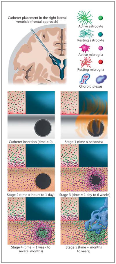
Although cerebrospinal fluid (CSF) shunt placement is the most common procedure performed by pediatric neurosurgeons, shunts remain among the most failure-prone life-sustaining medical devices implanted in modern medical practice. This article provides an overview of the mechanisms of CSF shunt failure for the 3 most commonly employed definitive CSF shunts in the practice of pediatric neurosurgery: ventriculoperitoneal, ventriculopleural, and ventriculoatrial. The text has been partitioned into the broad modes of shunt failure: obstruction, infection, mechanical shunt failure, overdrainage, and distal catheter site-specific failures. Clinical management strategies for the various modes of shunt failure are discussed as are research efforts directed towards reducing shunt complication rates. As it is unlikely that CSF shunting will become an obsolete procedure in the foreseeable future, it is incumbent on the pediatric neurosurgery community to maintain focused efforts to improve our understanding of and management strategies for shunt failure and shunt-related morbidity.

Keywords: Catheter obstruction; Hydrocephalus; Shunt failure; Ventriculoatrial shunt; Ventriculoperitoneal shunt; Ventriculopleural shunt.


Figures

Fig. 1
Fig. 1
Model of the parenchymal response to ventricular catheter placement and the most common form of noninfectious ventricular catheter obstruction. In the upper left, a coronal brain section containing a right frontal ventricular catheter is shown for reference. The box at the catheter entry point into the right lateral ventricle corresponds to the zoomed-in view of the subpanels below. In the upper right, a key to the cell types depicted in the model is provided. The subpanels correspond to the 5 stages described of the astrocyte/microglia centric model for ventricular catheter occlusion described in the text. The 1st subpanel, “catheter insertion” depicts the theoretical moment of catheter insertion prior to protein adsorption. In reality, as protein adsorption (“stage 1” sub-panel) occurs within microseconds of catheter placement, this process would actually be nearly complete by the time the surgeon has fully inserted the catheter. “Stage 2” depicts the initial tissue response to the implanted catheter, with microglia and astrocytes becoming activated and coalescing around the catheter shank within the brain parenchyma. The highly motile microglia serve as the leading front in this response and become most intimately associated with the portion of the catheter surface within the brain parenchyma. These microglia are the first to appear on the catheter surface in great numbers and, as they migrate along the catheter surface in a Brownian fashion, they adhere most readily to the irregular edges of the CSF intake holes and begin to accumulate at these sites in “stage 3.” After at least 1 week, if not more, in vivo astrocytes begin to appear in greater numbers, and, as depicted in “stage 4” can be seen forming cellular bridges spanning across the CSF intake holes. Over months to years the astrocytes begin to outnumber (or outcompete) the microglia. As depicted in “stage 5,” these astrocytes serve as a robust substrate for the secondary attachment of other cell types including choroid plexus (depicted) and sloughed ependymal cells (not depicted).
Fig. 2
Fig. 2
a–d Differential interference contrast images of transparent ventricular catheter illustrating imperfections on the surfaces of the CSF flow holes and the unevenness of the luminal surface. The images presented are of single optical sections illustrating approximately 2.5 μm in depth and were collected using a 3-dimensional, multispectral, spinning-disk confocal microscope (Olympus IX81 inverted microscope with motorized x-y-z stage, broad-spectrum light source, and charge-coupled device camera). Prior to imaging, the ventricular catheter was cut longitudinally, allowing a clear view of the irregular luminal and CSF intake hole surfaces.
Fig. 3
Fig. 3
Selected imaging illustrating distal catheter complications. a This chronically shunted teenage male presented with a 4-day history of abdominal pain and a 1-day history of headache associated with nausea and emesis. In this axial CT scan, the distal VP shunt catheter tubing is noted to be tightly coiled within a fluid collection contained within the intraperitoneal space, consistent with an abdominal pseudocyst. As is typical for abdominal pseudocysts, cultured samples of this fluid collection demonstrated the presence of Propionibacterium acnes. b This patient presented with new-onset headache and by radiographic shunt series was noted to have disconnection of his distal catheter at the site of a straight connector within the shunt system. c This patient present with localized, superficial abdominal swelling in the context of progressively worsening headache approximately 1 week after a distal catheter shunt revision. This coronal CT scan demonstrates distal VP shunt catheter tubing tightly coiled within a fluid collection contained within the preperitoneal space, most likely a reflection of suboptimal catheter placement at the time of the recent shunt revision surgery.

References

    1. Browd SR, Ragel BT, Gottfried ON, Kestle JR. Failure of cerebrospinal fluid shunts: part I: obstruction and mechanical failure. Pediatr Neurol. 2006;34:83–92. - PubMed
    1. Kestle J, Drake J, Milner R, Sainte-Rose C, Cinalli G, Boop F, Piatt J, Haines S, Schiff S, Cochrane D, Steinbok P, MacNeil N. Long-term follow-up data from the Shunt Design Trial. Pediatr Neurosurg. 2000;33:230–236. - PubMed
    1. Riva-Cambrin J, Kestle JR, Holubkov R, Butler J, Kulkarni AV, Drake J, Whitehead WE, Wellons JC, Shannon CN, Tamber MS, Limbrick DD, Rozzelle C, Browd SR, Simon TD, Network HCR. Risk factors for shunt malfunction in pediatric hydrocephalus: a multi-center prospective cohort study. J Neurosurg Pediatr. 2016;17:382–390. - PubMed
    1. Pollack IF, Albright AL, Adelson PD. A randomized, controlled study of a programmable shunt valve versus a conventional valve for patients with hydrocephalus. Hakim-Medos Investigator Group. Neurosurgery. 1999;45:1399–1408. discussion 1408–1311. - PubMed
    1. Scott RM, Madsen JR. Shunt technology: contemporary concepts and prospects. Clin Neurosurg. 2003;50:256–267. - PubMed