. 2017 May 15;77(10):2735-2745.
doi: 10.1158/0008-5472.CAN-16-1602. Epub 2017 Mar 1.

IL33 Promotes Colon Cancer Cell Stemness via JNK Activation and Macrophage Recruitment

Affiliations

IL33 Promotes Colon Cancer Cell Stemness via JNK Activation and Macrophage Recruitment

Min Fang et al. Cancer Res. .

Abstract

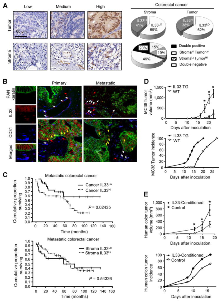
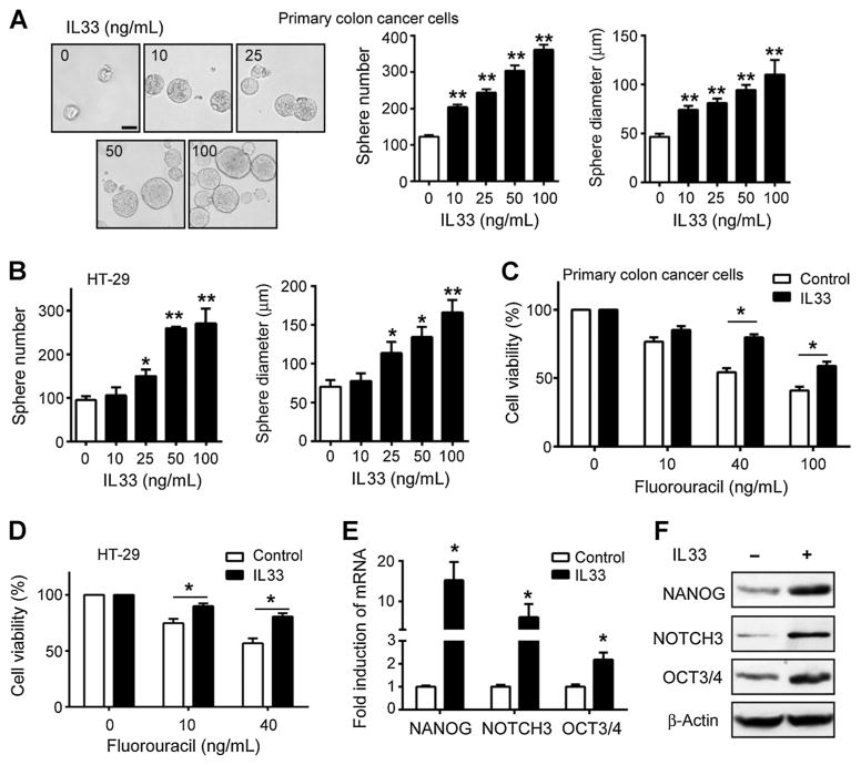
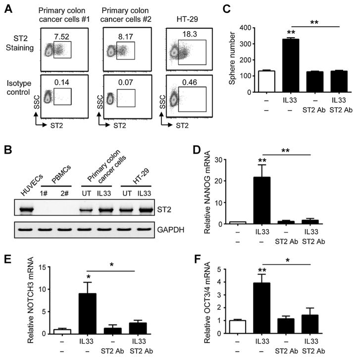
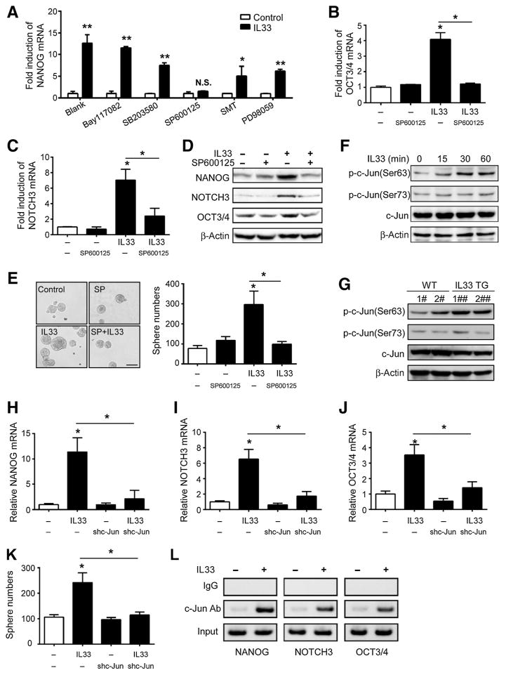
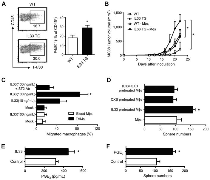
The expression and biological role of IL33 in colon cancer is poorly understood. In this study, we show that IL33 is expressed by vascular endothelial cells and tumor cells in the human colon cancer microenvironment. Administration of human IL33 and overexpression of murine IL33 enhanced human and murine colon cancer cell growth in vivo, respectively. IL33 stimulated cell sphere formation and prevented chemotherapy-induced tumor apoptosis. Mechanistically, IL33 activated core stem cell genes NANOG, NOTCH3, and OCT3/4 via the ST2 signaling pathway, and induced phosphorylation of c-Jun N terminal kinase (JNK) activation and enhanced binding of c-Jun to the promoters of the core stem cell genes. Moreover, IL33 recruited macrophages into the cancer microenvironment and stimulated them to produce prostaglandin E2, which supported colon cancer stemness and tumor growth. Clinically, tumor IL33 expression associated with poor survival in patients with metastatic colon cancer. Thus, IL33 dually targets tumor cells and macrophages and endows stem-like qualities to colon cancer cells to promote carcinogenesis. Collectively, our work reveals an immune-associated mechanism that extrinsically confers cancer cell stemness properties. Targeting the IL33 signaling pathway may offer an opportunity to treat patients with metastatic cancer. Cancer Res; 77(10); 2735-45. ©2017 AACR.


Conflict of interest statement

Disclosure of Potential Conflicts of Interest

No potential conflicts of interest were disclosed.

Figures

Figure 1
Figure 1
IL33 promotes colon tumorigenesis. A, IL33 expression was detected with conventional immunohistochemical staining in the human colon cancer tissues. The representative images of IL33 expression in colon cancer cells (Tumor) and stromal cells (Stroma) are shown. Scale bar, 60 μm. The proportion of IL33 expression in colon cancer cells and stromal cells in the colon cancer microenvironment is depicted (right, pie charts). B, IL33 expression was detected with multiplexed fluorescence staining in the human colon cancer tissues. The representative images show the expression of IL33 (red), CD31 (blue), PAN-Keratin (green). White arrows, nuclear IL33 localization in CD31+ vascular endothelial cells; yellow arrows, nuclear IL33 localization in Keratin+ tumor cells; red arrows, nuclear IL33 localization in CD31Keratin stromal cells. C, The association between the survival in patients with metastatic colon cancer and IL33 protein levels in tumor cells (top) and stromal cells (bottom). Survival functions were estimated by Kaplan–Meier methods and analyzed based on the H-score for tumor or stromal cell IL33 expression. D, MC38 cells (106) were subcutaneously injected into wild-type (WT) or IL33 transgenic (IL33 TG) mice. The tumor volume (top) and tumor incidence (bottom) were monitored. Results are expressed as the mean of tumor volume ± SEM; n = 7. *, P < 0.05. E, Human primary colorectal cancer cells (#1) were cultured with or without rhIL33 (0.1 μg/mL) for 24 hours. The cells (106) were subcutaneously injected into nude mice. The tumor volume (top) and tumor incidence (bottom) were monitored. Results are expressed as the mean of tumor volume ± SEM; n = 7, *, P < 0.05.
Figure 2
Figure 2
IL33 promotes colon cancer stemness. A and B, Effects of IL33 on colon cancer sphere formation. Primary colorectal cancer cells (A) and HT-29 cells (B; 4,000 cells) were subject to sphere assay for 7 days. IL33 was added on day 1 and 3. Representative images of spheres of primary colorectal cancer cells are shown (A, left). Scale bar, 50 μm. Results are expressed as the mean numbers of spheres ± SEM and the mean diameters of spheres ± SEM. Each column represents three independent experiments; n = 4; *, P < 0.05; **, P < 0.01. C and D, Effects of IL33 on colon cancer chemotherapy. Primary colorectal cancer cells (C) and HT-29 (D) were cultured with or without IL33 (100 ng/mL) for 24 hours and were subsequently exposed to 5-FU for 24 hours. The cell viability was determined by CCK-8 assay. Results are expressed as the mean ± SEM; n = 7; *, P < 0.05. E and F, Effects of IL33 on core stem cell gene expression in colon cancer cells. Primary colorectal cancer cells were treated with IL33 (100 ng/mL) for 24 hours. The levels of core stem cell gene transcripts (E) and proteins (F) were detected by real-time PCR and Western blotting, respectively. Each column represents three independent experiments. Results are expressed as the mean ± SEM; n = 4; *, P < 0.05.
Figure 3
Figure 3
IL33 promotes colon cancer stemness via its receptor ST2. A, Expression of ST2 protein in colon cancer cells. Primary colon cancer cells (#1 and #2) and HT-29 cells were stained with specific rabbit anti-human ST2 Ab and R-PE–conjugated goat anti-rabbit IgG. The expression of ST2 was determined by flow cytometer analyzer and expressed as the percentage of ST2+ cells in total colon cancer cells. One of four experiments is shown. B, Expression of ST2 mRNA in colon cancer cells. ST2 mRNA expression was determined by RT-PCR in human umbilical vascular endothelial cells (HUVEC), peripheral blood mononuclear cells (PBMC), primary colon cancer cells, and HT-29 cells. Primary colon cancer cells and HT-29 cells were cultured with 100 ng/mL IL33 for 24 hours. One of three experiments is shown. C, Effects of anti-ST2 on the role of IL33-mediated colon cancer sphere formation. Primary colorectal cancer cells were subject to sphere assay. IL33 (100 ng/mL) and/or anti-ST2 antibody (1 μg/mL) were added in the sphere culture. Results are expressed as the mean ± SEM; n = 4; **, P < 0.01. DF, Effects of anti-ST2 on the role of IL33-stimulated colon cancer stem cell gene expression. Primary colorectal cancer cells were cultured with IL33 (100 ng/mL) and/or anti-ST2 antibody (1 μg/mL) for 24 hours. The expression of NANOG (D), NOTCH3 (E), and OCT3/4 (F) transcripts were quantified by real-time PCR. Results are expressed as the mean ± SEM; n = 4; *, P < 0.05; **, P < 0.01.
Figure 4
Figure 4
IL33 promotes colon cancer stemness via c-Jun activation. AC, Quantification of NANOG (A), NOTCH3 (B), and OCT3/4 (C) transcripts in primary colorectal cancer cells that were treated with IL33 (100 ng/mL) for 24 hours in the presence of different signaling inhibitors (A) and the JNK inhibitor SP600125 (B and C). Each column represents three independent experiments. Results are expressed as the mean ± SEM; n = 4; *, P < 0.05; **, P < 0.01. D, Effects of the JNK inhibitor SP600125 on NANOG, NOTCH3, and OCT3/4 proteins in primary colorectal cancer cells. The cells were treated with IL33 (100 ng/mL) for 24 hours in the presence of SP600125 (10 μg/mL). NANOG, NOTCH3, and OCT3/4 proteins were detected by Western blotting. One of the three experiments is shown. E, Effects of SP600125 on colon cancer cell sphere formation. Primary colorectal cancer cells were subject to sphere assay in the presence of IL33 (100 ng/mL) and SP600125 (SP, 10 μg/mL). Representative images (left) and the mean numbers (right) of spheres are shown. Scale bar, 100 μm; n = 3; *, P < 0.05. F, Effects of IL33 on c-Jun in primary colorectal cancer cells. The cells were treated with IL33 (100 ng/mL) for different periods of time. The amount of phosphorylated c-Jun and c-Jun protein was detected by Western blotting. One of three experiments is shown. G, Phosphorylated c-Jun and c-Jun protein in MC38 tumors in vivo. MC38 tumor cells were obtained from wild-type and IL33 transgenic mice. The phosphorylated c-Jun and c-Jun proteins were detected by Western blotting. Representative data of two mice/group are shown. HJ, Effects of sh-c-Jun on IL33-stimulated stemness genes in primary colon cancer cells. Sh-c-Jun and scrambled vector expressing primary colorectal cancer cells were cultured with IL33 (100 ng/mL) for 24 hours. The mRNA levels of NANOG, NOTCH3, and OCT3/4 were detected by real-time PCR. Results are expressed as the mean ± SEM; n = 4; *, P < 0.05. K, The effects of sh-c-Jun on IL33-stimulated colon cancer cell sphere formation. Sh-c-Jun and scrambled vector expressing primary colorectal cancer cells were subject to sphere assay in the presence of IL33 (100 ng/mL). Sphere numbers were recorded. Results are expressed as the mean ± SEM; n = 4; *, P < 0.05. L, Effects of IL33 on the c-Jun occupancy in the promoters of core stem cell genes. Primary colorectal cancer cells were cultured with IL33 (100 ng/mL) for 24 hours. c-Jun-ChIP assay was performed. Rabbit IgG was used as a control. One of three experiments is shown.
Figure 5
Figure 5
IL33 promotes colon cancer stemness via recruiting and stimulating macrophages. A, Tumor-associated macrophages in IL33 transgenic mice. MC38 cells (106) were subcutaneously injected into wild type and IL33 transgenic mice. Tumor-infiltrating immune cells were stained for CD45 and F4/80 and were analyzed by FACS. Results are shown as the mean of F4/80+ macrophages ± SEM in CD45+ cells in day 28 tumor tissues; n = 4; *, P < 0.05. B, Effects of macrophage depletion on MC38 tumor growth. MC38 cells (106) were subcutaneously injected into wild type or IL33 transgenic mice. Clodronate liposomes were intraperitoneally injected into the mice. Tumor growth was monitored. Results are expressed as the mean of tumor volume ± SEM. – Møs, macrophage depletion; n = 4 per group; *, P < 0.05. C, Macrophage migration toward IL33. CD14+CD45+ macrophages were enriched and sorted from colon cancer tissues or normal blood and subjected to the migration assay in the presence of IL33 and/or anti-ST2. Results are expressed as the mean percentage of migrated cells ± SEM. Møs, macrophages. TAM, tumor associated macrophages; n = 4; *, P < 0.05. D, Effects of IL33-treated macrophages on colon cancer sphere formation. Normal blood CD14+ macrophages were treated with IL33 in the presence or absence of celecoxib for 72 hours. Primary colon cancer cells were subject to sphere formation in the presence of these treated macrophages. Results are expressed as the mean of sphere numbers ± SEM; n = 4 per group; *, P < 0.05. E, Effects of IL33 on macrophage-derived PGE2.Normal blood CD14+ macrophages were treated with IL33 for 48 hours. PGE2 was detected in the culture supernatants by ELISA. Results are expressed as the mean values ± SEM; n = 4 per group; *, P < 0.05. F, Effects of PGE2 on colon cancer sphere formation. Primary colon cancer cells were subject to sphere assay in the presence of PGE2 (50 ng/mL). Results are expressed as the mean of sphere numbers ± SEM; n = 4 per group; *, P < 0.01.

References

    1. Dinarello CA. An IL-1 family member requires caspase-1 processing and signals through the ST2 receptor. Immunity. 2005;23:461–2. - PubMed
    1. Schmitz J, Owyang A, Oldham E, Song Y, Murphy E, McClanahan TK, et al. IL-33, an interleukin-1-like cytokine that signals via the IL-1 receptor-related protein ST2 and induces T helper type 2-associated cytokines. Immunity. 2005;23:479–90. - PubMed
    1. Kurowska-Stolarska M, Kewin P, Murphy G, Russo RC, Stolarski B, Garcia CC, et al. IL-33 induces antigen-specific IL-5+ T cells and promotes allergic-induced airway inflammation independent of IL-4. J Immunol. 2008;181:4780–90. - PubMed
    1. Savinko T, Matikainen S, Saarialho-Kere U, Lehto M, Wang G, Lehtimaki S, et al. IL-33 and ST2 in atopic dermatitis: expression profiles and modulation by triggering factors. J Invest Dermatol. 2012;132:1392–400. - PubMed
    1. Tordesillas L, Goswami R, Benede S, Grishina G, Dunkin D, Jarvinen KM, et al. Skin exposure promotes a Th2-dependent sensitization to peanut allergens. J Clin Invest. 2014;124:4965–75. - PMC - PubMed

Publication types

MeSH terms

Substances