. 2017 May 1;77(9):2488-2499.
doi: 10.1158/0008-5472.CAN-16-2653. Epub 2017 Mar 1.

Kinome-Wide RNA Interference Screen Reveals a Role for PDK1 in Acquired Resistance to CDK4/6 Inhibition in ER-Positive Breast Cancer

Affiliations

Kinome-Wide RNA Interference Screen Reveals a Role for PDK1 in Acquired Resistance to CDK4/6 Inhibition in ER-Positive Breast Cancer

Valerie M Jansen et al. Cancer Res. .

Erratum in

Abstract

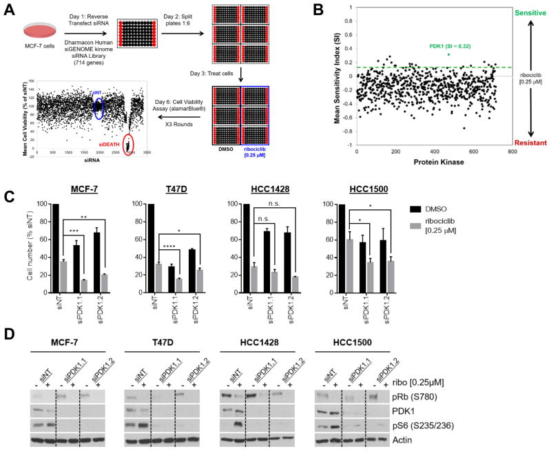
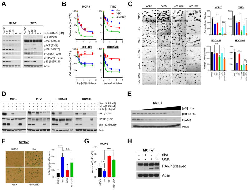
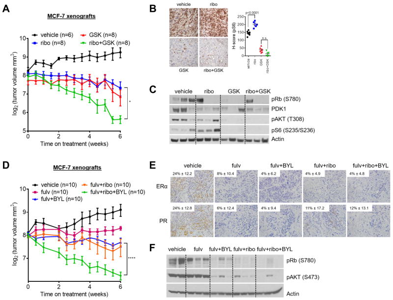
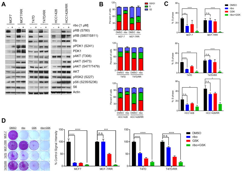
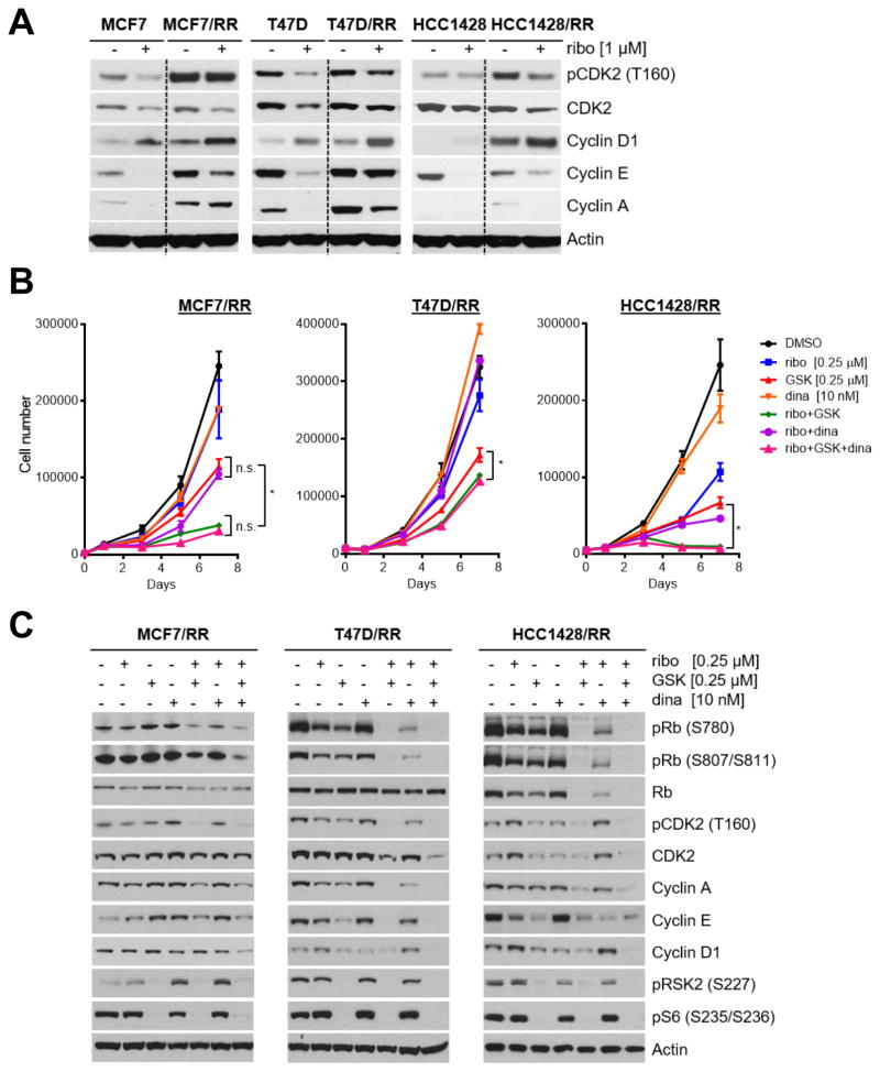
Acquired resistance to cyclin-dependent kinases 4 and 6 (CDK4/6) small-molecule inhibitors in breast cancer arises through mechanisms that are yet uncharacterized. In this study, we used a kinome-wide siRNA screen to identify kinases that, when downregulated, yield sensitivity to the CDK4/6 inhibitor ribociclib. In this manner, we identified 3-phosphoinositide-dependent protein kinase 1 (PDK1) as a key modifier of ribociclib sensitivity in estrogen receptor-positive MCF-7 breast cancer cells. Pharmacologic inhibition of PDK1 with GSK2334470 in combination with ribociclib or palbociclib, another CDK4/6 inhibitor, synergistically inhibited proliferation and increased apoptosis in a panel of ER-positive breast cancer cell lines. Ribociclib-resistant breast cancer cells selected by chronic drug exposure displayed a relative increase in the levels of PDK1 and activation of the AKT pathway. Analysis of these cells revealed that CDK4/6 inhibition failed to induce cell-cycle arrest or senescence. Mechanistic investigations showed that resistant cells coordinately upregulated expression of cyclins A, E, and D1, activated phospho-CDK2, and phospho-S477/T479 AKT. Treatment with GSK2334470 or the CDK2 inhibitor dinaciclib was sufficient to reverse these events and to restore the sensitivity of ribociclib-resistant cells to CDK4/6 inhibitors. Ribociclib, in combination with GSK2334470 or the PI3Kα inhibitor alpelisib, decreased xenograft tumor growth more potently than each drug alone. Taken together, our results highlight a role for the PI3K-PDK1 signaling pathway in mediating acquired resistance to CDK4/6 inhibitors. Cancer Res; 77(9); 2488-99. ©2017 AACR.


Conflict of interest statement

Conflict of Interest: The authors disclose no potential conflicts of interest.

Figures

Figure 1
Figure 1. Kinase screen identifies PDK1 siRNA as sensitizer to CDK4/6 inhibitor
(A) Overview of high-throughput screening (HTS) method. MCF-7 cells were reverse transfected with siRNA in 96-well plates. Each plate contained 80 individual siRNAs, indicated in black, and supplemented with controls, indicated in red [no siRNA, non-targeting control siRNA (siNT), and siDEATH positive control]. Transfected cells were divided into 6 replicate plates. Half of the plates (n=3) were treated with DMSO (vehicle control) and half (n=3) with 0.25 μM of ribociclib. Cell viability was assessed after 72 h of drug exposure using the AlamarBlue® reagent (Invitrogen). The experiment was repeated 3 times. (B) Scatter plot of the mean sensitivity index (SI) scores for 714 protein kinases and kinase-related proteins averaged across the three screening trials. A cutoff SI >0.15 (indicated by dotted line) was used for hit selection. The position of PDK1 (SI score 0.32) is noted. (C) ER+ MCF-7, T47D, HCC1428 and HCC1500 breast cancer cell lines were transfected with one of two siRNAs targeting PDK1 (siPDK1.1 and siPDK1.2), and a non-targeting control siRNA (siNT) and treated with DMSO (vehicle control) or 0.25 μM ribociclib for 72 h. Knockdown of PDK1 decreased cell proliferation and this effect was enhanced upon simultaneous treatment with ribociclib. (D) Immunoblot analyses of the cells following PDK1 knockdown and treatment for 72 h with DMSO or 0.25 μM ribociclib. Experiments were performed in full serum condition in the presence of endogenous estrogen (in IMEM/10% FBS/0.002% Phenol red).
Figure 2
Figure 2. Combined PDK1 and CDK4/6 inhibition reduces ER+ breast cancer cell proliferation
(A) ER+ MCF-7 and T47D cells were treated for 24 h with DMSO or increasing concentrations of GSK2334470. Lysates were prepared for immunoblot analysis with the indicated antibodies. GSK2334470 treatment diminished the expression of downstream targets of PDK1. (B) Cells were treated with DMSO or increasing concentrations of ribociclib, GSK2334470, or the combination for 7–10 days, after which nuclei were stained with DAPI and counted on the Molecular Devices ImageXpress instrument. Data are presented as the percent of cells remaining compared to the control (CTL, DMSO-treated). In all cell lines, combined inhibition of PDK1 and CDK4/6 was most effective than each drug alone. (C) Cells seeded in Matrigel were treated with DMSO or 0.25 μM ribociclib ± 0.25 μM GSK2334470. After 10–21 days, colonies were stained with the MTT reagent, photographed (left) and counted (right) using the GelCount® reader. In MCF-7 and T47D cells, specifically, combined inhibition of PDK1 and CDK4/6 was more effective than single-agent inhibition. (D) Cells were treated with DMSO, 0.25 μM ribociclib ± 0.25 μM GSK2334470, or 0.25 μM palbociclib ± 0.25 μM GSK2334470 for 24 h, after which lysates were prepared for immunoblot analysis with the indicated antibodies. Only combined inhibition of PDK1 and CDK4/6 led to concomitant decreases in P-Rb and P-S6. (E) MCF-7 cells were treated for 72 h with DMSO or increasing concentrations of ribociclib. Immunoblot analysis of the lysates showed that ribociclib decreased P-Rb and FoxM1 levels. (F) MCF-7 cells were treated with DMSO or 1 μM ribociclib ± 1 μM GSK2334470 for 72 h and analyzed for senescence by β-galactosidase staining. Ribociclib alone or in combination with GSK2334470 induced senescence compared to DMSO-treated cells. Data represent the average percent of senescence-associated (SA)-β-galactosidase positive cells per 5 high-power fields. (G) MCF-7 cells were treated with DMSO or 1 μM ribociclib ± 1 μM GSK2334470 for 72 h, stained with annexin V and propidium iodide, and analyzed by FACS. GSK2334470 alone or in combination with ribociclib increased the percent of apoptotic cells compared to DMSO-treated cells. (H) MCF-7 cells were treated with DMSO or 1 μM ribociclib ± 1 μM GSK2334470 for 72 h. Immunoblot analysis of these lysates revealed PARP cleavage only when cells were treated with GSK2334470 alone or in combination with ribociclib. Unless noted, media and drugs were replenished every 2–3 days (**, P < 0.01; ****, P < 0.0001 by ANOVA).
Figure 3
Figure 3. Pharmacological inhibition of PI3K/PDK1 enhances the effect of ribociclib in vivo
(A) MCF-7 cells were injected s.c. into athymic ovariectomized female mice, each supplemented with a short-term, 14-day release 17β-estradiol pellet. Mice bearing tumors ≥150 mm3 were randomized to vehicle, ribociclib, GSK2334470 or the combination of ribociclib and GSK2334470 for 6 weeks. Data are presented as log2 of mean tumor volume in mm3 (*, P < 0.05 vs. single-agent ribociclib or GSK2334470). Numbers in parenthesis represent the number of mice per treatment arm. (B) Representative images of tumor sections from A and quantitative comparison of P-S6 histoscores (H-score). GSK2334470 ± ribociclib inhibited P-S6; single agent ribociclib increased P-S6 levels. (C) Xenografts from A were homogenized after the last dose of drug treatment and tumor lysates were subjected to immunoblot analysis for the indicated antibodies. (D) MCF-7 cells were injected into mice as in A. Mice bearing tumors ≥150 mm3 were randomized to vehicle, fulvestrant, BYL719 and fulvestrant, ribociclib and fulvestrant, or fulvestrant, BYL719, and ribociclib for 6 weeks. The triple combination was most effective at decreasing tumor volume compared to single-agent therapy or double-combinations. Data are presented as log2 of mean tumor volume in mm3 (****, P < 0.0001 vs. fulvestrant, fulvestrant and BYL719, or fulvestrant and ribociclib). (E) Representative images of tumor sections from D and quantitative comparison of ER and PR histoscores (H-score) confirming target inhibition with fulvestrant. (F) Xenografts from D were homogenized after the last dose of drug treatment and tumor lysates were subjected to immunoblot analysis for the indicated antibodies.
Figure 4
Figure 4. CDK4/6 inhibition increases PDK1 expression in ER+ breast cancer cells and in primary tumor explants
(A) MCF-7 cells were treated with ribociclib over a time course up to 72 h. Cell lysates were prepared and subjected to immunoblot analyses with the indicated antibodies as described in the Methods. (B) Patient tumor explants were treated with DMSO or palbociclib for 48 h. Representative IHC for PDK1, S235/236 P-S6 and cyclin D1 is shown. Tumor explant 2 exhibited high basal levels of PDK1 and P-S6, while explants 1 & 3 exhibited drug-induced increases in PDK1, P-S6, and cyclin D1 levels. (C) PDK1, P-S6, and cyclin D1 IHC analysis of serial primary tumor sections from two patients before treatment and on the 7th day of treatment with palbociclib.
Figure 5
Figure 5. PDK1 inhibition restores sensitivity to CDK4/6 blockade in drug-resistant cells
(A) Lysates from parental MCF-7, T47D, HCC1428 and ribociclib-resistant cells treated ± 1 μM ribociclib were analyzed by immunoblot with the indicated antibodies. Resistant cells were removed from drug for 24 h prior to ribociclib treatment for this analysis. (B) Parental and ribociclib-resistant (RR) cells were serum starved for 24 h, treated with 1 μM ribociclib for 24 h, stained with propidium iodide, and then analyzed by FACS. (C) Cells were serum starved for 24 h as in A, treated with drugs for 24 h, stained with propidium iodide, and then analyzed by FACS. In all cases, combined treatment with ribociclib and GSK2334470 markedly reduced the percent of cells in S-phase (*, P ≤ 0.05; **, P ≤ 0.01; ****, P ≤ 0.0001). (D) MCF-7 and T47D parental and RR cells were seeded at low density in 12-well plates and treated with fresh media and drugs every 2–3 days. Resistant cells were removed from ribociclib for 24 h prior to drug treatment. Cells were treated with 1 μM of each drug in all experiments. After 10–21 days, cell monolayers were stained with crystal violet and subjected to image analysis as indicated in the Methods. Each bar represents the mean image signal intensity ± SD of triplicate wells (***, P ≤ 0.001; ****, P ≤ 0.0001).
Figure 6
Figure 6. Expression of cell cycle cyclins and CDKs is sustained in ribociclib-resistant cells
(A) MCF-7, T47D, HCC1428 parental and ribociclib-resistant (RR) cells were treated ± ribociclib for 24 h. Cell lysates were then prepared and analyzed by immunoblot with the indicated antibodies. (B) Cells were plated in triplicate in 12-well plates and treated with ribociclib, GSK2334470 and dinaciclib alone or in combination as indicated. Cells were trypsinized and counted on days 0, 1, 3, 5, and 7 of treatment. Media and drugs were replenished on days 3 and 5. Ribociclib/GSK2334470 or the triple combination of ribociclib/GSK2334470/dinaciclib was most effective at inhibiting cell growth compared to single agent or ribociclib/dinaciclib (* P ≤ 0.05). (C) RR cells were treated with ribociclib, GSK2334470, dinaciclib or combinations of these drugs for 24 h; cell lysates were analyzed by immunoblot for the indicated proteins. Resistant cells were removed from ribociclib for 24 h prior to drug treatment. The combination of ribociclib/GSK2334470 or the triple combination was most effective at diminishing expression of cyclins and CDKs.

References

    1. Finn RS, Crown JP, Lang I, Boer K, Bondarenko IM, Kulyk SO, et al. The cyclin-dependent kinase 4/6 inhibitor palbociclib in combination with letrozole versus letrozole alone as first-line treatment of oestrogen receptor-positive, HER2-negative, advanced breast cancer (PALOMA-1/TRIO-18): a randomised phase 2 study. Lancet Oncol. 2015;16:25–35. - PubMed
    1. Infante JR, Shapiro G, Witteveen P, Gerecitano JF, Ribrag V, Chugh R, et al. A phase I study of the single-agent CDK4/6 inhibitor LEE011 in pts with advanced solid tumors and lymphomas. Journal of Clinical Oncology. 2014:32.
    1. Shapiro G, Rosen LS, Tolcher AW, Goldman JW, Gandhi L, Papadopoulos KP, et al. A first-in-human phase I study of the CDK4/6 inhibitor, LY2835219, for patients with advanced cancer. Journal of Clinical Oncology. 2013:31.
    1. Turner NC, Huang Bartlett C, Cristofanilli M. Palbociclib in Hormone-Receptor-Positive Advanced Breast Cancer. N Engl J Med. 2015;373:1672–3. - PubMed
    1. Patnaik A, Rosen LS, Tolaney SM, Tolcher AW, Goldman JW, Gandhi L, et al. Efficacy and Safety of Abemaciclib, an Inhibitor of CDK4 and CDK6, for Patients with Breast Cancer, Non-Small Cell Lung Cancer, and Other Solid Tumors. Cancer discovery. 2016;6:740–53. - PubMed

MeSH terms